95 Matching Annotations
  1. Sep 2016
    1. peculiar morphogenesis with alteration of proliferation patterns and of conserved signaling pathwa

      Scales that cover the feet of birds have been shown to be different to other scales, with different molecules and development patterns, so it could be independently developed.

    2. It has been previously hypothesized (47) that reptilian scales are more similar to avian reticulate scales (covering the foot pad) than to both avian scutate scales (covering the anterior metatarsal region) and feathers

      The scales on the feet pads of birds (e.g. chickens) show very similar pathways to the ones in reptiles, far into the development of the embryo.

      In contrast, this similarity is not as easily observable in feather or foot covering scales compared to reptile scale development, leading to works hypothesising that they are not related.

    3. feathers are organized into discrete tracts associated to different body areas

      The development of feathers develop in 10 different areas and these areas correspond to different parts of the body.

      These tracts develop at different rates and have their own patterning.

    4. Bmp4 is also shown for lizard. Red double-headed arrows indicate the body region processed for sectioning.

      the authors have also managed to demonstrate the expression of Bmp4 in lizard, which strongly suggests a developmental evolutionary link between all reptiles, and birds and mammals.

    5. caudal
    6. spinal
    7. cervical
    8. ventral

      the underside or the abdominal part of the body https://upload.wikimedia.org/wikipedia/commons/6/64/Horse_Axes.JPG

    9. immunohistochemistry

      The process for detecting antigens in tissues by using the principle that antibodies binding specifically to antigens.

      And this method is used in the recognition of abnormal cells, and for visualising the distribution and localization of specific cellular components.

    10. humeral

      an anatomical term which refers to a bone called humerus

      https://upload.wikimedia.org/wikipedia/commons/e/e4/Humerus_-_anterior_view.png

    11. femoral
    12. (A and B) WMISH with Ctnnb1 in C. niloticus and P. vitticepsembryos at various developmental stages. Arrowheads indicate the initiation sites of scale tracts and arrows indicate the directions of scale tracts. Colors correspond to different tracts schematically represented in the right panels (dots, initiation sites; arrows, directions of development). (C) WMISH with Shh in P. guttatus embryos at various developmental stages. Arrowheads with white borders indicate tract initiation sites, and arrowheads with black borders indicate the boundaries of Shh expression at different developmental stages, showing the different anteroposterior (a/p) and ventrodorsal (v/d) gradients (see schematic in the right panel).

      the authors claim that anatomical placodes in reptiles have been overlooked in previous studies; here they demonstrate them in reptiles by ISH with genes that corporate in conserved signalling pathways that are involved in skin patterning and early morphogenesis. as a result the authors prove the importance of anatomical placodes in reptile is not less than other amniotes.

    13. lateral
    14. mutant scaleless chicken
    15. Hematoxylin and eosin (H&E)

      Histologists- those who study tissues- dye different parts of cells prior to studying them under microscope.

      H&E is a widely used staining, which makes nuclei blue and the plasma pink, respectively.

    16. sebocytes

      any kind of cell that secretes sebum- an oily substance which contains fat-

    17. nested subpopulation
    18. integument

      the outer layer that mainly serves as protection

    19. keratinized

      to change to a form that contains keratin- a fibrous protein which mostly found in hair, nail, hoof, etc.-

    20. whole-mount in situ hybridization (WMISH) with species-specific probes, we show that crocodile, lizard, and snake placodes all exhibit spatial expression of Shh

      The WMISH experiments (used to detect gene expression) show that Shh, a morphogen needed in development, was expressed in the placode.

    21. superposed

      superposing means to put or lay something over something else

    22. These results indicate that all three characteristics (epidermal thickening, expression of epidermal placode markers, and expression of dermal Bmp4), that is, the presence of an anatomical placode, are required for proper development of scales in reptiles.

      the presence of an anatomical placode, are required for proper development of scales in reptiles. as shown all the molecular markers of anatomical placode presents in the sca; just the same as wild type. but due to the inability of sca to form anatomical placode because of another mutation; they fail to form scales properly.

    23. histological

      pertaining to histology-the science of studying tissues-

    24. Similarly, Ctnnb1 andEdar, two other placode markers, also show marked differences in expression between wild-type and scaleless dragons. In both phenotypes, expression of these two genes is first ubiquitous across the whole epidermis before becoming restricted to the placodes in wild-type individuals only (Fig. 4B). These results indicate that expression of each of these three placode markers in reptiles is similar to the expression dynamic of the corresponding genes in mammals (27) and birds (20, 28,39). On the other hand, the absence of an anatomical placode in scaleless dragons coincides with the inability of signaling pathways to pattern the skin, similar to what is observed in mice deficient in Eda/Edar(40).

      the authors are demonstrating that not only the signalling pathway but also the anatomical placode are required for reptiles to develop scales properly.

    25. generates a new splice donor site (gt) 42 bases upstream of the wild-type donor site, thus generating a 14–amino acid deletion in the corresponding transcript (Fig. 3D).

      The insertion of the jumping DNA caused a mutation. This mutation affected splicing, a process where the DNA is edited to ultimately produce the correct protein.

      This incorrect splicing resulted in a shortened non-functional protein.

    26. in-frame deletion

      As you know from molecular biology, protein-coding region of DNA transcribe into mRNAs and then, mRNAs, in their turn, translate into proteins on ribosomes. Basically three nucleotides are required to code for any aminoacid, so the part of mRNA that translates into proteins has a "frame" which is divisible by three; if a deletion cause removal of three or a multiplicate of three then the frame will stop at the previous stop codon and other aminoiacs remain untouched, otherwise the deletion may turn the protein to anything at all.

      https://upload.wikimedia.org/wikipedia/commons/thumb/6/6d/RNA-codons-aminoacids.svg/2000px-RNA-codons-aminoacids.svg.png

    27. Fig. 3

      TAB ONE - Panel A

      A picture taken from above of the wild-type lizard and the scaleless mutant.

      The white arrows show the large spines protruding from the sides on the wild-type individual.

      In contrast the Scaleless mutant doesn't have any scales protruding.

      TAB TWO - Panel B

      A picture taken from below of the wild-type lizard and the scaleless mutant.

      The white arrows show the position of secretary glands surrounded by rows of scales that stick out from the inner thigh of the wild-type.

      The absence of arrows show that these glands are not found in the Scaleless mutant.

      TAB THREE - Micro X-Ray images METHOD

      Micros-rays use electromagnetic radiation which give them a high penetrating imaging system. This allows the inside of objects and living organisms to be imaged. This is done at a microscopic level, allowing very small elements to be imaged, such as in this case, where a lizard skull and ‘hand’ are imaged

      Computed tomography takes the combination of multiple x-ray images taken at different angles to then create a cross sectional ( a slice) image, as seen in Panel C.

      TAB THREE - Panel C

      X-ray images of the skull and 'hands'.

      The white frames on the skulls show the position of the pleurodont ( regenerating lizard teeth).The wild-type show normal teeth while the scaleless mutant have smaller and fewer teeth.

      The double-headed arrows show the length of the claws. The wild-type can be seen to have shorter claws than the scaleless mutants.

      TAB FOUR - Panel D

      Upper panel

      The image shows a simplified representation of the EDA protein (which is made up of amino acids).

      The EDA protein is made up two distinct conserved areas- a collagen region and a TNF (tumour necrosis factor). the ares highlighted in red is the most conserved region.

      In the wild-type, all the domains are found to be intact.

      In the scaleless mutant, 15 amino acids are missing from the extremely conserved area in the TNF domain, leaving only 3 amino acids of the high conserved area.

      Lower Panel

      The highly conserved area (displayed as a red area in the higher panel) is represented in red letters. This area is compared in different species (mouse, chicken, pogona lizard).

      This sequence is found in all the species except in the mutant. Because it is found across many different species, this proves that is highly conserved. this usually indicates that it plays an important function.

      TAB FIVE - Splicing Process

      DNA is assembled in a code so that genetic information can be stored. Genes which are made up of DNA, act as individual units of information. The DNA of a gene will be ‘read’ and turned into RNA, which act as a messenger. This information will then be ‘read’ to form proteins. Proteins are functional and can perform actions and functions e.g. causing reactions, degrading etc.

      However each gene is highly processed in a process called splicing. This process will edit the RNA; intron regions (non functional/ non coding regions) of a gene are removed and exons (regions that are read) are linked together.

      Incorrect splicing can lead to a non-functional protein.

      TAB 6 - RT PCR

      Reverse transcription polymerase chain reaction

      TAB 7 - Panel E

      The image above shows a representation of the structure the EDA gene at specific area. The area represented is composed of Exons 7 & 8 and the intron between these two exons.

      The length of the intron that is removed is indicated (1.2 kb). the donor site (gt) and acceptor site (ag) are the sites where the DNa is cut and these two sites are then joined together.

      Primers used in RT-PCR are indicated in blue: F1, F2, R1 and R2.

      In this experiment, the splicing outcomes are verified and compared between the wild-type and the scaleless mutant. This is done using RT-PCR with the primers indicated with blue arrows: F1, F2, R1 and R2.

      The results indicate that there are large differences between the wild-type and the mutant. This is due to the presence of a transposon in Exon 7, outlines in red in the image on the top.

      TAB SEVEN - Panel F

      The cells of the wild-type’s back (dorsal) and front (lateral), shown in the two leftside images, were stained and compared to the same stained cells of the mutant, shown on the two right side images.

      Three photos were taken of each place of each lizard

      On the top row, H and E staining

      The wild-type, show protruding skin sections, indicating presence of scales

      ON the middle row. Immuno-flourescent staining was used.

      This highlighted the proteins: α-keratins (α-k) and β-keratins (β-k) or laminin. Keratin is a protein found in scales whereas laminin is found on the outer layers of skin. The wild-type has thick beta keratin layer which is almost entirely missing in the mutant. The laminins show a weird formation in the mutants, as they are much more bumpy.

      ON the last row, Toluidine blue (TB) staining was used on the dorsum (back)and scanning electron microscopy (SEM) was used on skin molts;

      TB staining shows a clear scale outline in the wild-type whilst it is completely flat in the mutant. The skin molts clearly have different textures between the wild-type and mutant.

      TAB EIGHT - Panel G

      The wild-type lizard (P. vitticeps) embryo at different stages of development (24, 28, 38, 44 days after beginning of development). The image in the corner show the overall embryo with a red arrow indicating where the image of the cell stained image was taken. The cell staining images show when the scale starts to form.

      TAB NINE - Conclusion Overall, these images show the difference between the wild-type lizard and the mutant lizard. The mutant has no scales (panel A and F), not as many teetch and longer claws (panel B and C). It also has a an inserted transposon in the gene (panel E) leading to a mutated EDA protein (panel D).

    28. homologous

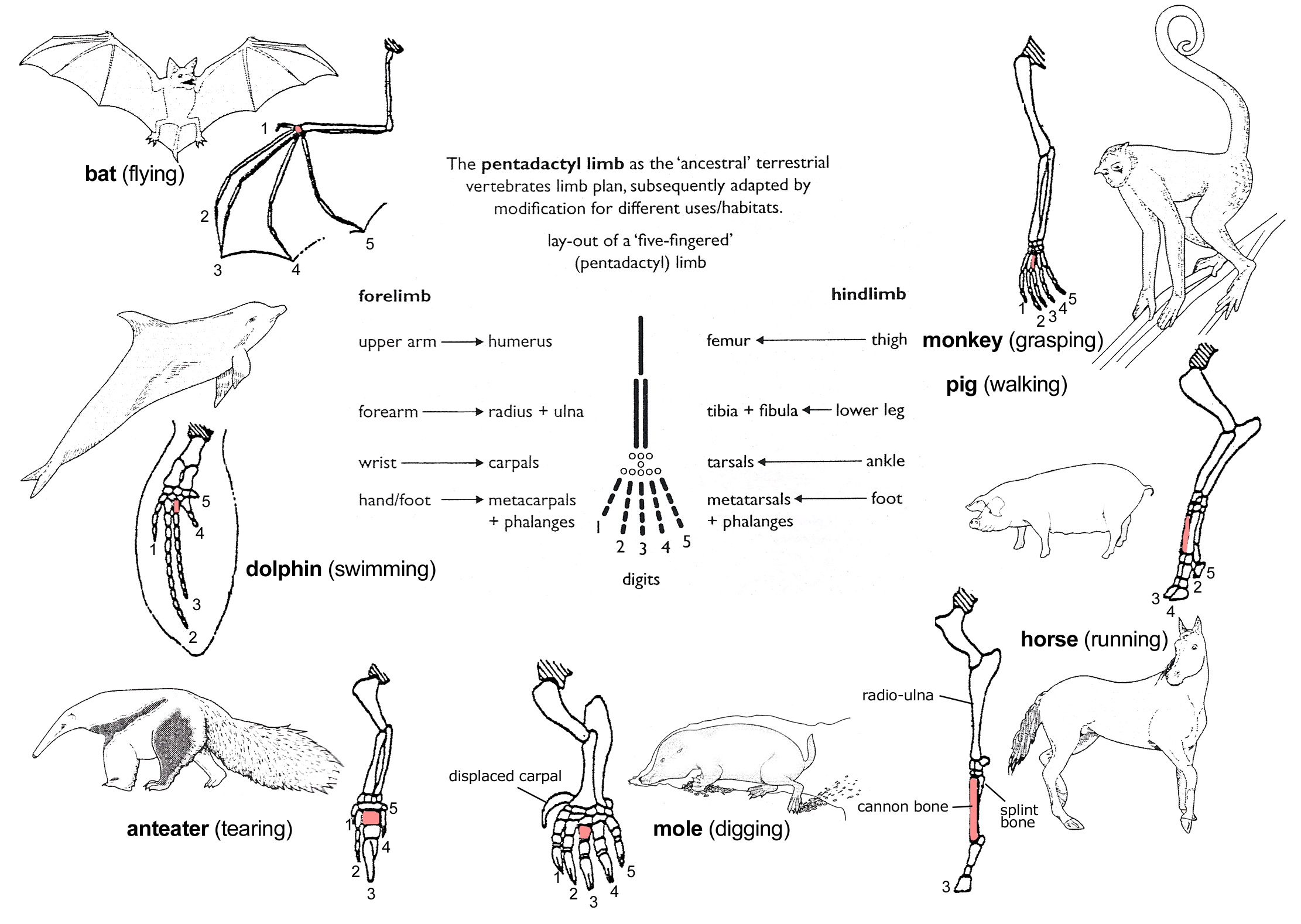
      homologous properties in biology have the same evolutionary origins but not necessarily a same function. like human and bat's arm.

      https://upload.wikimedia.org/wikipedia/commons/5/5b/Evolution_pl.png

    29. Using breeding experiments,

      The authors took different individual lizards with different physical traits, and then mated them to see how these traits were transmitted to the offspring of these mixes.

    30. crocodiles
    31. reptiles
    32. spatiotemporal development is highly similar between the two species

      when you say spatiotemporal- space-time- development of macropatterning tracts are similar; you mean that these macropatterning tracts develop in a similar space and at a similar timepoint during the developmental process.

    33. scutate

      covered with scutate-A horny or bony external plate.!

      https://upload.wikimedia.org/wikipedia/commons/1/17/Alligator_foot_detail.jpg

    34. lineage

      A sequence of species that have evolved from a same ancestor.

      a sequence of species that have evolved from a same ancestor.

      https://upload.wikimedia.org/wikipedia/en/6/68/Diagram_of_Eusocial_Species.png

    35. cryosections

      sections that are made in a cryostat- a device for keeping the temperature low-.

    36. PCNA

      A protein of DNA polymerase delta. it is involved in the control of eukaryotic DNA replication. PCNA is important for replication and participates in cell division. so it's present in cell shows that the cell is proliferating.

    37. Anatomical placodes in C. niloticus (left panels), P.vitticeps (middle panels), and P. guttatus (right panels) embryos. For each species, the whole-embryo WMISH with Sonic hedgehog (Shh) is shown (left panel) as well as, from top to bottom, high magnification of H&E-stained placode sections (white arrowheads indicate placode columnar cells), immunohistochemistry with PCNA (proliferation marker; epidermal-dermal junction indicated by dashed white lines), and parasagittal cryosections of placodes after Shh or β-catenin (Ctnnb1) WMISH. Bmp4 is also shown for lizard. Red double-headed arrows indicate the body region processed for sectioning.

      expression of the Sonic hedgehog (Shh), β-catenin (Ctnnb1), genes, are amongst the conserved molecular markers of anatomical placodes. Here, the author proves that, indeed, these genes are being expressed at morphogenetic-anatomical placode- cells in reptiles, by ISH. You may also see the development of scales in all shown spices at different developmental stages. note that in all cases scale development starts from the formation of anatomical placodes, just like other amniotes.

    38. parasagittal

      an imaginary plane that divides the body into right and left halves.

    39. (A) Hematoxylin and eosin (H&E) staining of skin sections from different body regions (indicated with red arrows on the top insets with lateral views of corresponding embryos) of C. niloticus (crocodile; top row), P.vitticeps (lizard; two middle rows), and P. guttatus (snake; bottom row) embryos at various developmental stages [indicated as embryonic days (E) after oviposition]. White arrowheads indicate the anatomical placode. Scale bars, 100 μm.

      The author is trying to show the anatomical placode, using H&E staining. as shown, anathmical placodes are generally like a thickening. another characteristic of anatomical placodes is reduced proliferation. note that as you see in figure B, in placodes the proliferation marker (PCNA) does not exist, but in nearby the white arrow, the small green dots represent PCNA, then, proliferation.

    40. Abstract 

      Are feathers, hairs, and scales family?

    41. EDA (ectodysplasin A)

      This is a protein involved in cell signalling between two layers of skin (ectoderm and mesoderm) , which is especially important in embryo formation.

      This allows the formation of hair follicles, sweat glands, and teeth.

    42. INTRODUCTION

      Introduction

      Do skin appendages like hair in mammals, feathers in birds, and scales in reptiles come from the same origin? Due to substantial differences in their look and some differences in the genes they express, this question has confused scientists for long time. This article tries to settle this controversy by its new findings in the development of scales in reptiles, and shows that we may all be more closely related than we thought.

    43. Conversely, other authors argue that the similarities in molecular signaling observed among all skin appendages suffice to support their homology (9). Note that such placodal signaling centers have been recently evidenced as underlying the development of Chelonia shell scutes

      Other works have contradicted models where there is independent evolution of these traits.

      They suggest that due to the similarity of the molecules in the processes, they are closely related and come from a common ancestor.

      It is also suggested that this is true for turtles as well which have been shown to need the placode for the development of their shell.

    44. Both models assume that the development of an anatomical placode and of a dermal papilla occurred, at a minimum, twice (once in birds and once in mammals) through the independent parallel co-option of the same set of signaling pathways (WNTs, β-catenin, EDAR, BMPs, and SHH)

      The two models proposed by various papers over time both propose that the evolution of the placode occurred independently twice.

      This means that the signalling molecules were evolved independently in different organisms but ended up with similar functions.

    45. Finally, similar in situ hybridization analyses indicate the presence and total absence of Bmp4 dermal expression in wild-type and scaleless dragons, respectively

      ISH (in situ hybridisation) was used to show that the expression of the BMP4 gene.

      It was found that it was expressed in the wild type but not the mutant.

    46. The most marked and derived macropatterning of skin in reptiles is observed in snakes

      The features of snake skin developed was visualised with WMISH, showing that snakes have a unique pattern of development of scales.

    47. Our WMISH experiments, with early developmental scale markers, such as Shh and Ctnnb1, on developmental series of Nile crocodiles and bearded dragon lizards clearly indicate

      WMISH (Whole-mount in situ hybridization) was the experiment technique used. This is a technique which labels RNA using fluorescence etc.

      RNA is produced by genes that are being expressed so by seeing what RNA there is, we can see what genes are being used.

      In this study, WMISH was used to detect RNA of developmental genes (e.g. Shh, Ctnnb1) in crocodiles and bearded dragon lizards.

    48. BMP

      A group of chemicals that are growth factors.

    49. indicate that early scale morphogenesis in reptiles consists of regular dermoepidermal elevations that typically further develop into oriented asymmetrical scales with various levels of overlap, depending on the species and body area

      Scales form with risen areas in the dermo-epidermal layer (the skin) which then further develop into scales which are different depending on the species and where it is on the body.

    50. proliferating cell nuclear antigen (PCNA) analyses indicate a reduced proliferation rate of the placode epidermal cells

      proliferating cell nuclear antigen (PCNA) is a protein which is necessarry in replication, and therefore if it is detected, this means the cells will be reproducing.

      this analysis showed that the cells of the placode were reproducing very slowly.

    51. Our serial sectioning and histological analyses of skin developmental series (Fig. 1A) in crocodiles (Crocodylus niloticus), bearded dragon lizards (P. vitticeps), and corn snakes

      Serial sectioning is successive microscopic images of histological- which means tissues. Here, microscopic images were taken of different body parts of the lizard and and snakes, focussing on where scales form.

    52. argued homologous to those characterized in chicken

      Because the same patterns in development of chicken feathers and lizard scales are similar, it can be assumed that they are related.

    53. This set of new results coherently and conclusively indicates that most skin appendages in amniotes are homologous; that is, they all evolved from a shared common ancestor that exhibited appendages developing from an anatomical placode and expressing a set of signaling molecules still involved in the development of scales, hairs, and feathers of extant species.

      The placode is crucial in the formation of all the different skin appendages (hair, feathers, scales), and all of them develop with the same molecules.

      This shows that they are homologous- thus closely related.

    54. similarities in signaling are due to independent co-option of these molecular pathways.

      Previous work suggests that although there are similar molecules found in the development of feathers, hair and scales, they all function differently and evolved independently.

    55. L. Alibardi, Adaptation to the land: The skin of reptiles in comparison to that of amphibians and endotherm amniotes. J. Exp. Zool. B Mol. Dev. Evol. 298, 12–41 (2003).

      this review focusses on the adaptation of skin of reptiles as they evolved from water-dwelling amphibians to

    56. R. B. Widelitz, T.-X. Jiang, J. Lu, C.-M. Chuong, b-catenin in epithelial morphogenesis: Conversion of part of avian foot scales into feather buds with a mutated b-catenin. Dev. Biol. 219, 98–114 (2000).

      This paper explored the role of β-catenin (protein involved in growth and thought to be a morphogen) which is expressed in the placode.

      It was found that when this protein was mutated, the chickens would be scaleless(on the feet) and have abnormal feather growth.

    57. C. Blanpain, E. Fuchs, Epidermal stem cells of the skin. Annu. Rev. Cell Dev. Biol. 22, 339–373 (2006).

      The development of skin cells that give rise to hairs is reviewed in this paper.

      A hair placode forms which allow for the expression of genes that determine skin cells and hair follicle development.

    58. D. Dhouailly, A new scenario for the evolutionary origin of hair, feather, and avian scales. J. Anat. 214, 587–606 (2009).

      This paper proposed the theory that mammal hairs evolved from glandular structures. Whereas reptiles and birds skin (including feathers and scales) evolved a different pathway where a thick protective covering which would become scales.

    59. P. F. A.Maderson,Mammalian skin evolution: A reevaluation. Exp. Dermatol. 12, 233–236 (2003).

      This review deals with a model of how hair evolved, updating a model built in the 1972 (reference 3).

      This model supposes that the development of hair was caused by mutations in patterning genes. this development allowed hair to become useful insulation.

    60. M. C. Milinkovitch, L. Manukyan, A. Debry, N. Di-Poï, S. Martin, D. Singh, D. Lambert, M. Zwicker, Crocodile head scales are not developmental units but emerge from physical cracking. Science 339, 78–81 (2013).

      This paper illustrates the way that crocodile head scales form. the process is different to the formation of other skin traits (such as feathers, hair and other scales).

      The crocodile face and jaw scales are formed through the force of the growing cells physically pushing and cracking about hard shell forming unique patterns.

    61. we show for the first time

      To read a popular science version of this paper, check out motherelode's article, complete with quotes from the author.

      http://motherboard.vice.com/read/hairy-humans-and-scaly-lizards-have-more-in-common-than-you-think

    62. reveal a new evolutionary scenario

      http://www.sciencemag.org/news/2008/11/so-thats-why-chickens-have-combs

      It has also been previously suggested that proteins involved in hair formation are found in reptiles for claw formation and in birds for beak formation. This hinted at the shared ancestry of hair, claws and scales.

    63. J. M. Musser, G. P. Wagner, R. O. Prum, Nuclear b-catenin localization supports homology of feathers, avian scutate scales, and alligator scales in early development. Evol. Dev. 17, 185–194 (2015).

      The relationship of feathers and scales are explored in this research paper.

      The location and amount of a morphogen ( a chemical which causes/allows for development of traits), Beta-catenin is visualised during development, showing very similar patterns. This led the author to conclude that they both evolved from the same thing.

    64. P. F. A. Maderson, When? Why? and How?: Some speculations on evolution of vertebrate integument. Am. Zool. 12, 159–171 (1972)

      Review from the 1970’s discussing the formation of integuments (hard protective layer covering the body), scales, hair. The age of this review gives an insight into previous hypotheses and the historical research on this matter.

      The different hypothesis of the time are explored including; scales being a precursor to non scaled animals, ossified (boney) scales being a precursor to scales found on current reptiles and the evolution of hair from sensory appendages.

    65. A. M. Turing, The chemical basis of morphogenesis. Philos. Trans. R. Soc. London Ser. B 237, 37–72 (1952).

      Turing's landmark paper where he put forward the reaction diffusion theory to explain morphogenesis.

      This theory states that two evenly distributed chemicals interact with each other, and by inhibiting and activating each other, then rearrange to form chemical gradients which can then form patterns.

    66. discrete developmental units through reaction-diffusion

      Morphogens (chemicals which allow formation of traits), interact with each other to form specific concentrations at certain areas to form patterns and determine the placing of appendages.

      This is done by the chemicals activating and repressing the chemicals around them to varying degree leading to unique chemical gradients

    67. dermoepidermal elevations

      An area of tissue that joins the epidermal (outer layer) and the dermal layers (middle layer) of the skin.

      Elevations refer to bumps found in this layer, leading to areas that are slightly raised.

      https://upload.wikimedia.org/wikipedia/commons/e/ec/Anatomy_of_the_skin.jpg

    68. columnar cells

      Cell's shape that are similar to the columns and it's height is at least four times their width.

      https://upload.wikimedia.org/wikipedia/commons/c/c5/Simple_columnar_epithelial_cells.png

    69. common ancestry

      On of the principles of darwin's Evolutionary theory that says organisms share a most recent common ancestor.

      https://upload.wikimedia.org/wikipedia/commons/thumb/7/70/Phylogenetic_tree.svg/2000px-Phylogenetic_tree.svg.png

    70. parasagittal

      An imaginary plane that divides the body into right and left halves.

    71. nonconcurrently

      Not happening at the same time

    72. signaling pathways

      Refers to a group of molecules in a cell that work together to control one or more cell functions.

    73. (TNF)

      The tumor necrosis factor (TNF) superfamily : a group of cytokines that take place in apoptosis.(cell death)

    74. codominant

      Two alleles of a gene in a heterozygote that are both fully expressed.

    75. pleurodont

      Tooth fused to the inner edge of the jaw.

      This means it is loosely attached and can regenerate if it is lost.

    76. autopod

      futherest part of the limb, such as the hand or foot.

    77. These results reveal a new evolutionary scenario where hairs, feathers, and scales of extant species are homologous structures inherited, with modification, from their shared reptilian ancestor’s skin appendages already characterized by an anatomical placode and associated signaling molecules.

      The authors have found that the placode and the placode's signalling molecules are highly conserved - meaning it is found across many species.

      This study shows that the placode is responsible for the formation of many different structures in the various species, such as feathers, scales and hair.

    78. co-option

      A structure or system with an original function evolves so that the it adds or changes to a new function.

    79. Several studies (9, 18–23) have shown that conserved signaling pathways, evidenced by the expression of the Sonic hedgehog (Shh), β-catenin (Ctnnb1), ectodysplasin A receptor (Edar), and/or bone morphogenetic protein (Bmp) genes, are involved in skin patterning and early morphogenesis of all amniote skin appendages

      The same signalling pathways were found in various different amniotes, suggesting it is a conserved across many species, and thus important.

      These include:

      Sonic Hedgehog (Shh)- factor in formation of organs and appendages.

      β-catenin (Ctnnb1) - Factor in gene expression and cell-cell adhesion.

      Ectodysplasin A receptor (Edar)- Factor in skin and nervous system development.

      Bone morphogenetic protein (Bmp)-Factor in skeletal formation.

    80. material cracking

      Due to exertion of force, the living material physically cracks the scale tissues. This leads to unique patterns on each crocodilian face.

    81. follicular organs

      A small spherical group of cells containing a cavity in which materials are contained and can grow.

      Example: hair, teeth, feathers

    82. homology

      In biology, similarity of the structure, physiology, or development of different species of organisms based upon their descent from a common evolutionary ancestor.

    83. molecular markers

      Basically, in biology each cellular function achieves by corporation of protein complexes; for example in mammals, DNA replication happens by DNA polymerase and a dozen of other molecules( for example PCNA); so if we recognise any of these molecules that make DNA replication happens; it shows that the cell is proliferating;

    84. wild-type

      A type of the typical form of a species in which seen in nature.

    85. avian

      Relating to, or characteristic of birds.

    86. placode

      A platelike structure, especially a thickening of the ectoderm marking the site of future development in the early embryo

    87. squamates

      The order Squamata, or the scaled reptiles, are the largest recent order of reptiles, comprising all lizards and snakes.

    88. morphogenesis

      A biological process in which embryo develops to adult in some organisms.

    89. fossil intermediate

      A transitional fossile that gives us information about a transition from one species to another. Actually is one that falls between "before transition forms" and "after transition forms"

    90. homozygous for a codominant mutation

      Homozygous means that they have two copies of the exact same gene.

      Co-dominant means that two different genes that are present in the same individual will both be expressed and blended together.

      In this case, the gene which causes a scaleless mutant must have two copies of the mutant gene. If there is one copy, there will be partial formation of scales.

    91. Extant

      Life form that is currently in existence.

      Antonym: extinct

    92. similar phenotypes in other vertebrates because of impairments of the EDA receptor (EDAR; a member of the TNF family) (18) or its ligand EDA, indicating a conserved role of this pathway in reptiles as well

      Similar disease symptoms can be observed in other animals with a mutation in the same gene.

    93. Scaleless dragons show an irregular skin surface with the initiation of some dermoepidermal undulations of the skin (Fig. 3G), indicating that this phenomenon does not fully require the presence of anatomical placodes.

      Even though the mutant does not have scales as a result of a non functional protein, it still shows the beginnings of a pattern for scales.

      This means that there are other elements that are not part of the anatomical placode which contribute to the formation of scales

    94. cDNA

      DNA (which stores genetic information) makes RNA (which transports genetic information) which is then read to make a protein which is functional

      cDNA is a copy of the RNA which is tells us what protein will be created.

    95. ectodermal dysplasia syndrome

      A genetic disease which affects the growth of hair, teeth, nails and sweat glands.