Raúl Magario, responsable del Área de Logística y de Finanzas,
Logistica y Finanzas de Montoneros
Raúl Magario, responsable del Área de Logística y de Finanzas,
Logistica y Finanzas de Montoneros
Communicate, It depends how you feel when you are around people. Some people have angry or happy. They got bad vibe.
studiousness, which has nothing to do with hitting the books. “Studiositas” is sort of like cultivated attention, like training that desire to know so that it’s focused on the good stuff as opposed to looking at TikTok for five hours a day.
And we forget that the Greek word, the root for “school,” is leisure.But leisure is not idleness or amusement. And it’s definitely not just resting up so you can get back to work. It is that space that we need to set aside to cultivate the highest parts of us.
leisure vs. entertainment vs. work vs. occupation vs. labor
But I think there’s a remarkable kind of red thread running through all of that, which is this idea that there is something really essential and important, not just to individuals, but to culture and society, in having something that is more than an education that we would call professional, and that they would call servile.
let’s just take Great Books education for example. You find Great Books education in community colleges. You find Great Books education, still, completely outside of institutions of higher education. You find Great Books education in certain high schools.I think these things don’t necessarily have to be luxury goods. And I think it’s a choice that we make, politically, to say that they are. We can debate that choice, but that’s just a choice that we have.
If you’re going to read a Dan Brown novel, it’s very difficult to imagine having sustained conversations about Dan Brown novels over years. It’s quite easy to imagine doing that with Shakespeare. I do that. It’s just so rich.And so I think we should not shy away from saying that. There is a kind of depth in great art that demands our attention in a way that is absent in Dan Brown.
that cultivating the life of the mind is a basic human need.
but it’s really striking to read about the role that the liberal arts played in these large-scale working class communities in the past.
Is Douthat aware of the broader background and playing a bit of devil's advocate here?
Douthat: But then isn’t there an argument, a critique of the humanities, that says that intellectual mentality and the eugenic mentality could fit together pretty naturally? It’s like: OK, to be human is to appreciate Bach and Plato, and only our smartest university students do that, so only they’re fully human — and so on down the eugenicist argument.
Douthat buys into the idea that you have to be "smart" to appreciate the humanities or the great books.
Didn't Mortimer J. Adler spend a large chunk of his life trying to convince "everyman" that they could appreciate Plato and Aristotle?
When you look at a culture, and you want to ask yourself: “Well, how did we go from Weimar Germany to Nazism?” Obviously, education is going to be a part of that, but it’s not in any way going to be the whole of it.
One of the same front, Catholics seem to be big in America behind the Great Books and classical education, yet also seem to be a large group of conservatives behind MAGA/Trumpism.
you also have to recognize that there is an ineliminable element of human freedom in education.
Nazis despite a humanistic education....
Is the link that Douthat a real one?
1692
fear, social tension, and personal grudges fueled the witchcraft accusations.
This shows how the gaspesian man pushed back against French assumptions by explaining that his community's way of life was based on balance, respect, and freedom not "savagery" he forced the reader to see native society on its own way.
I believe the message shows how the puritans believed their new colony a moral responsibility. His city on a hill idea explains why they felt their success or failure would be seen by the world.
The irony is that most Wall Street people freak out about high Federal budget deficits and debt, but those actually produce safe securities, namely treasury bonds. When Wall Street generates private debt, like mortgages and commercial credit, that’s what fosters financial crisis.
Good that he notes this.
o make values comparable across counties with different assessment cycles and price levels, we express each property’s value as a percentile rank across all 1–4 family residential parcels in the study area
percentiling doesn't actually help with making things comparable across counties. percentiling mostly helps because assessed values in general trail market values, so we care more about relative assessed than absolute assessed
Discussion
my thoughts on the outline of this section: - pinch point recap, opportunity to increase funding + meet more demand - one para on lower-value, less-white - one para on repeat vs. not; perhaps targeting could factor in a property's flood history more than just if they flooded really badly in one event (which is how most HMGP works) - what we learn about mechanisms - limitations in methods - limitations conceptually in terms of not knowing if what we see is "good for people" or not
).
there is no discussion in here about the repeat-flooded panel. it's notable tha the share applying is still small, but at least is double the share in the "all-flooded" group. Also notable that their success rate in applying is about the same as in the all-flooded group.
and we discuss any sensitivity of our results to this distinction in the Supplementary Materials.
flagging since we do not currently do this
the Repetitive Flood Claims Program (RFC), and the Severe Repetitive Loss Grant Program (SRL)
is FMA the combination of RFC and SRL?
This property-resolved flood history is, to our knowledge, the first parcel-scale, multi-event reconstruction of repetitive flooding in the region, and serves as the reference set against which subsequent pipeline stages are measured.
well, helena's own paper does this for buildings, so I'm not sure this is really true
located
I don't know where this should go ,but i think we should acknowledge that we don't know if this distribution is equitable in that we don't know if people are better off after being mitigated than before (especially in the case of buyouts). i would want to head off people reading this and going "oh ok everything looks fine" when there could be elements of coercion into applying into buyouts + ending up worse off than before. So I think we want to frame it as "we can rule out some procedural issues, but we're not saying that just because lower-value properties are getting the money, there are no issues"
Neighborhood
similar to my comment above, i would compress this paragraph a lot and maybe even combine it with the one above.
The neighborhood-scale selection we observe could reflect several mechanisms: cost-benefit analysis criteria that favor lower-cost properties, programmatic concentration of buyouts to enable contiguous open-space conversion, or differential outreach by local governments to particular neighborhoods. Disentangling these is an important direction for future work.
I think we should have a stand-alone paragraph that addresses what we DO learn about mechanisms from this analysis. we see no evidence that the CBA is skewing selection to HIGHER value properties, and in theory that should apply across states (since it's a federal rule). We also don't see evidence that a bunch of communities are just choosing to not apply - but the state match could have a lot to do with that. It doesn't look like the communities are only going to expensive properties and asking them to apply, otherwise we'd see a more expensive applicant pool.
Among the small fraction of properties that do receive funding, the composition shifts toward less expensive properties and lower-value neighborhoods. Funded properties sit at the th percentile of the study-area property-value distribution, compared to the th percentile of all flooded properties. The shift occurs primarily between neighborhoods rather than within them: within-block-group percentiles for funded properties differ only modestly from those at the flooded stage, indicating that funded properties tend to be typical homes in lower-value neighborhoods rather than the cheaper homes within any given neighborhood.
this feels like too much text to dedicate to recapping results; let's compress
This replicates at parcel resolution what the prior literature has documented at the county scale.
delete, duplicative of above
applicants
One aspect it'd be good to bring in here is that the mitigations have been repeatedly found to be cost-effective, so expanding the funding could likely translate to more cost-effective mitigations -- basically, there are good reasons to expand the pool, not just make the pool more competitive
While
because the long methods are at the end, i think we need one paragraph at the start of results that describes briefly what we actually did, so that the jump into results isn't so abrupt.
A map of our data (apps, mits, the study area) would also be good to include i think
eligible
need to remove language around "eligible" throughout
fall disproportionately on lower-income households in under-resourced communities
I'm not sure what this means. the requirements apply to everyone, so how can they "fall disproportionately" on some households? it's possible that it can be a more difficult challenge to meet for some households, e.g. heirs property issues, being able to be at home at the time the inspector shows up, etc. But I think we would need a citation for that specifically.
.
Insert text here about the 25% match. something like "providing the 25% non-federal share can also be a barrier. in some states, the state government covers the matching share. If not, the funding has to come from local government budgets or individual homeowners, which can significantly constrain participation"
North Carolina
rephrase this generally as we haven't started talking about NC at all yet. "states, local governments, and in some cases homeowners are responsible for the remaining 25% non-federal match."
A
1) i would move this paragraph down to the next section where we describe all the possible reasons why disparities might arise. we already have a line on CBA at the end, so let's just put it all in one place
2) I would call this a potential driver of the disparity -- we don't actually know for sure this is happening. projects are also cheaper when they are for lower-value homes, so the cost side goes down in addition to the benefit side. Also, there are "exemptions" to the CBA rule -- things like "if an elevation costs less than X and the property was substantially damaged, it passes". So i'd frame it more as CBA might be one driver but that's not known.
Concerns about equitable access to FEMA assistance have been documented in the immediate aftermath of disasters for decades.
Pretty sure this is my sentence, but revise to: Concerns about equitable access to FEMA assistance in the aftermath of disasters have been extensively documented
The
Suggest restructuring this paragraph the fed gov't is the primary source of public assistance for disaster risk mitigation and post-disaster relief and recovery. Many federal agencies are involved in these efforts, including XYZ. We focus our analysis on FEMA...
is it true that FEMA's mitigation spending > USACE? Probably depends on how you define it
that are not being selected
replace with "applying and not being selected"
,
insert "grant programs" after (HMA)
, which bear a disproportionate flood burden,
i think this can be deleted, otherwise we need a specific citation (one that gets at distribution of impacts not just hazard)
in FEMA hazard mitigation grants alone
i think you can delete. not worth introducing the whole concept of FEMA hazard mitigation grants
projected climate changes will
adjust to "climate change is projected to intensify..."
more than twice the figure suggested by FEMA’s official flood maps,
suggest deleting
Patients B
Case#: Family 2: Patient B, a 10 y/o girl of Indian origin
DiseaseAssertion: Guanidinoacetate methyltransferase deficiency. Pathogenic homozygous missense variant in exon 5 of GAMT gene.
FamilyInfo: Born to nonconsanguineous parents. Uneventful birth history. Parents were confirmed to be carriers.
CasePresentingHPOs: HP:0002078, HP:0001249, HP:0040082, HP:0001263, HP:0001344, HP:0002474, HP:0002360, HP:0000752, HP:0000252
CaseHPOFreeText: Global developmental delay first was noted at 8–10 months of age when they had not achieved neck holding and social smile. They had no speech, poor expressive language, and cognition. They also had hyperactivity, sleep disturbances, frequent bursts of laughter, and happy predisposition. Reverse phenotyping with MRS brain confirmed an absent creatine peak in both siblings. The blood creatinine in patient B was 0.18 mg/dl (0.015 mmol/L), which is low.
CaseNotHPOs: HP:0032794, HP:0008542, HP:0000572, HP:0001999, HP:0031422
CaseNotHPOFreeText: No hearing or vision loss. On examination of both siblings, there was no dysmorphism. The cranial nerve assessment, tone, and deep tendon reflexes were normal. There were no abnormal cerebellar signs except for truncal ataxia. The MRI brain, EEG, and CMA were normal. The blood tandem mass spectrometry and urine GC-MS were normal. The methylation-specific PCR study for Angelman syndrome was normal.
usually demonstrated by direct comparison with available related software
What an we compare this to
Katie J. Wells quote from near the end of the interview:
...when you have very very low expectations for public government, Silicon Valley looks like an OK alternative... the technology in your pocket somehow looks more useful.
This says so much about citizenship and the relationship between democracy, autocracy, and technology.
tasks
Quiz Missing
In
Missing H2 from original -
Welcome to Module #!
Was this deleted intentionally?
For the specific grading criteria for this assignment, review the rubric at the bottom of this page.
We don't want to say bottom of the page per wcag.
Change to:
Review the specific grading criteria for this assignment in the rubric.
instruction
instructions
Select the Prepare tab for preparation information, the Complete tab for assignment instructions, the Submit tab for submission requirements, and the Evaluation tab for grading information.
Add [Add overview] before instructions
Change language to:
Select Prepare for preparation information, Complete for assignment instructions, Submit for submission requirements, and Evaluation for grading information.
It could always turn out that one of the objections is instantly recognized by the entire group as a fatal flaw in "foo" and the group will then turn to a discussion of the merits (and demerits) of "bar" instead.
Getting everyone to agree about what are the negative properties of each proposal is key. Without judgement. The engineering is then about figuring out if the negatives of proposal bar are worse than the negatives of foo. Often it's not the better proposal that wins, but the proposal that has fewest (or no) showstopping negatives..
— m
You're using em dashes too much.
a result that a credible public test strictly helps authors whose default standing sits below the bar — and we are precise about the downside it carries for those just above it;
The language of this is a bit unclear. Try to make it easier to understand.
(1) a
Don't use bold here.
r to a public-evaluation venue that pays expert evaluators
I'm not sure if the fact that evaluators are paid here is relevant to this question - rather than giving these details, you could just say "The Unjournal is the focal example"
he Post tab for detailed instructions on how to make your initial post, the Respond tab to review the requirements for responding to your peers, and the Evaluation tab for information on how this discussion will be graded.
This was changed from the language we provided, which doesn't include "the" and "tab".
Change to:
Select Post for instructions on your initial post, Respond for peer response requirements, and Evaluation for grading information.
please, consult the Discussion Rubric.
Change to:
please consult the rubric.
remove extra comma, "Discussion", and bold
Select
Add [Add overview] before instructions
please, consult the Discussion Rubric.
Change to:
please consult the rubric.
remove extra comma, "Discussion", and bold
Select the Prepare tab to review key content necessary for participating in the discussion, the Post tab for detailed instructions on how to make your initial post, the Respond tab to review the requirements for responding to your peers, and the Evaluation tab for information on how this discussion will be graded.
Add [Add overview] before instructions
Change language to:
Select Prepare to review key content for the discussion, Post for instructions on your initial post, Respond for peer response requirements, and Evaluation for grading information.
Slang refers to new or adapted words that are specific to a group, context, and/or time period; regarded as less formal; and representative of people’s creative play with language.
This is exactly like playing Arma Reforger online with my friends. We make up random tactical words that change all the time depending on the situation. If you are new to the server you probably wont understand anything we say. Its just a fast way for us to talk during the game.
Communicating emotions using “I language” may also facilitate emotion sharing by not making our conversational partner feel at fault or defensive.
Group projects can facilitate this kind of stuff. If I tell someone they never help they more than likely get mad and do even less. If I say I am feeling stressed about the work and I could use the help they usually step up.
One feature of communicative incivility is polarizing language, which refers to language that presents people, ideas, or situations as polar opposites. Such language exaggerates differences and overgeneralizes.
You see this everywhere on Facebook. People act like everything is completely black or white and they pick a side. Seems the focus is to create division and no one actually listens to each other. It basically just shuts down the whole conversation and wastes time. This has lead to me ridding my phone of the app. Except for marketplace nothing better then looking at cool stuff I cannot afford.
Większość ludzi biega ŹLE. Jak trenować po 40-tce? Adam Kszczot
code-switching refers to changes in accent, dialect, or language (Martin & Nakayama, 2010). There are many reasons that people might code-switch. Regarding accents, some people hire vocal coaches or speech-language pathologists to help them alter their accent
I do this at my IT help desk job on campus all the time. I talk totally normal to other students when they come in for help. But when my boss or a teacher comes in I switch up and sound super professional.
The triangle of meaning is a model of communication that indicates the relationship among a thought, symbol, and referent and highlights the indirect relationship between the symbol and referent
This makes sense when I think about talking with my wife about house stuff. We use the same word but picture completely different things in our heads. It usually leads to a dumb argument over nothing.
lema anterior, t
Citar H2
Lema.
H2
Lema.
H1
f
$f$
constannte
constante
Bolzado-
Bolzano
Weierstrass(citar en preliminares),
Falta
con los siguientes resultados
previamente con la validez de los siguientes resultados
resécto
respecto
desigualdad maximal de Doob
Añadir a preliminares
ultimo,
último
es completamente valida
está bien definida.
(Se moverá a preliminares)
Falta
Always read the selection at least twice, no matter how long it is.
I definitely have had to learn to do this over the last few years. I used to read quickly through something and did not understand I needed to go back and re-read if things were not sinking in for me.
An Archive of a Different Type<br /> by [[Jason Scott]] on Internet Archive Blogs accessed on 2026-06-11T10:58:48
Hanging from a wall near Martin and his bowtie and lab coat was a sign reading “Psychoanalysis For Your Typewriter.”
Imagine being so well-known for your craft that letters addressed to “Mr. Typewriter, New York” would get delivered by the Post Office to your door.
Around the mid-19th century, Swiss astronomer Rudolf Wolf rolled a pair of dice 20000 times, recording each outcome.
wtaf
Ron's Typewriter - YouTube<br /> by [[Parks and Recreation]] <br /> accessed on 2025-10-06T22:09:41
Parks and Recreation S3, E5 "Media Blitz" aired 2011-02-17<br /> https://www.imdb.com/title/tt1636241/
Can you think of any words not included in this list that would be helpful to know in relationship to your college vocabulary?
I think that counseling would be helpful to know in your college vocabulary.
When and where might you need each of the words?
You will need to use these words while you are in school.
Will you need some words more frequently than others?
I do think that you will need to use some words more than others.
Will you need all the words all the time?
You don't need these words all the time, but it can be helpful to know what they mean.
Are there any connections between the words that you can see?
The words are all connected because they relate to college and academics.
Can you see any ways to simplify the task of learning 30 words?
You can use repetition to help simplify learning 30 words where you can go over them multiple times.
Major
A field of study that someone specializes in.
Learning Community
A community of people that can help each other learn.
Co-requisite
I do not know what this is.
Prerequisite
Something you need to do before other courses.
Upper Division Course
A higher level class.
Lower Division Course
A lower level and easier class.
Pre-College Level Course
A course at a pre-college level.
College Level Course
A course that is at college level.
Course Number
A number to identify a certain course.
Grade Options
Different ways for a student to be graded.
Audit
I do not know what an audit is.
Non-Credit/Continuing Education
A class that doesn't give you credits.
Transcript
A document of a students grades.
Federal Work Study
Work that the school can give you.
Grant
Money given to a student to help them pay for school.
Scholarship
Money that is given to a student for college that has excelled in something.
Are gamers goal oriented?
Gamers are goal oriented because they play games where the goal is to beat certain levels or build up to reach their goal.
eLife Assessment
This important study provides evidence that plateau pikas, at moderate densities, can facilitate yak nutrition by suppressing a poisonous plant, offering a helpful perspective on reciprocal interactions between small mammal ecosystem engineers and large herbivores. The evidence is solid, supported by a manipulative field experiment and appropriate measurements of intermediary ecological processes, although some claims about density dependence, competition, and stress-gradient mechanisms are not fully supported by the experimental design. The work will be of interest to ecologists, conservation biologists, and rangeland managers, particularly those studying grassland herbivore interactions and livestock management on the Qinghai-Tibetan Plateau.
Reviewer #1 (Public review):
Summary:
This is important and significant work because it helps describe the complexity of interactions between system components where two herbivores interact with vegetation. Whereas other studies have shown that the larger ungulate (yaks, Bos grunniens, in this case) can facilitate the abundance and population growth of the smaller (the semi-fossorial lagomorph, Ochotona curzoniae, plateau pika hereafter), this study flips the tables and shows that, at least under some conditions, moderate densities of the plateau facilitate the nutritional condition of yaks.
The study was not designed to investigate the reasons that pikas clip Stellera chamaejasme. That said, based on other studies and general knowledge of the ecology of these pikas, it is likely that they clip (although do not eat) this plant because its relatively large size hinders predator detection. This species of pika does better where vegetation height is low than where it is higher.
Strengths:
Notably, the strong inference the authors can claim for their results is supported by the careful experimental design. A weaker paper would have simply noted correlations between pika burrow density and yak feeding efficiency without experimental removal. This paper, to its credit, not only used experimental removals but also documented the various intermediary results that support the ultimate conclusions. The statistical approaches used appear to be appropriate. (Readers are encouraged to read the full Materials and Methods, which are available in the Supplementary Materials section.)
Weaknesses:
Although the study was well designed and executed, and its conclusions appear strongly supported, readers interested in the management implications of the Qinghai-Tibetan Plateau should be mindful of its limitations. First, the study site, at approximately 3,200 m elevation, was relatively low by Qinghai-Tibetan Plateau standards. Stellera chamaejasme becomes less common at elevations > 4,000 m, where a majority of livestock grazing occurs. Thus, it would be instructive to learn, through follow-up studies, whether similar facilitation occurs where unpalatable (and mildly poisonous) species in such genera as Astragalus, Oxytropis, and Thermopsis replace S. chamaejasme as the problematic plant for pastoralists. Second, the authors make no mention of wild ungulates, so it is unclear what, if any, role they may have played in this system. At least one study in Qinghai Province, albeit at a slightly higher elevation, showed that not only pikas, but also Tibetan gazelles (Procapra picticaudata), which were commonly observed on grazed pastures, grazed more frequently on some dicots avoided by domestic sheep than did the livestock themselves (Harris et al. 2015). It would also be instructive to learn if similar facilitation as observed here applied to the other principal livestock species in the area, domestic sheep (which are often herded together with smaller numbers of domestic goats). Finally, as suggested by this study, the interactions between all components of the system are complex and interactive. If pika facilitation of yak nutrition at the densities documented results in herders increasing yak density, might the increased herbivory from the domestic animals provide the conditions for the pika population to increase beyond the densities observed here, and thus toward the levels where facilitation yields to competition?
Citation:
Harris RB, Wang, WY, Badinqiuying , Smith AT, Bedunah DJ (2015) Herbivory and Competition of Tibetan Steppe Vegetation in Winter Pasture: Effects of Livestock Exclosure and Plateau Pika Reduction. PLoS ONE 10(7): e0132897. doi:10.1371/journal.pone.0132897
Reviewer #2 (Public review):
Summary:
This study uses a combination of field sampling and manipulative experiments to test for facilitative impacts of pikas on yaks via suppression of a poisonous forb. The authors found that, when Stellera forbs were present, yak weight increases over the growing season were greater in the presence of pikas compared to in their absence. This occurred because, although pikas do not consume Stellera, they clip it and use it in nest/burrow construction, thereby decreasing its relative abundance in the plant community. Thus, overall, the study contributes to our understanding of how herbivores of different size classes indirectly affect each other via the use of shared resources.
Strengths:
It is well known that large herbivores on grasslands impact smaller animals, but the reciprocal interaction is rarely tested. Thus, this study asks a valuable question, and the experiment is well-designed to test it. The authors also do a good job of demonstrating the potential conservation impacts of their research.
Weaknesses:
What the authors tested is really cool, but their claims go far beyond what they can say based on their experimental design. For example, the authors claim to show that pika impacts on yaks display density-dependent transitions from competition to facilitation. However, their experiment only looked at the presence (at moderate densities) and absence of pikas, and they only tested for facilitation, not competition.
The paper would also benefit from changes to the framing in the introduction and discussion. For example, the authors pitch the work as a test of the stress-gradient hypothesis. However, there is no abiotic stress gradient in the study, which is an essential component of the SGH. They also pitch the work in terms of density dependence, but there is no significant variation in population densities beyond the presence-absence binary. The paper would be stronger if they focused their framing around the literature on facilitative interactions across mammals of different size classes, especially indirect facilitation via use of shared resources, which is what this paper is really about.
Finally, the paper has significant weaknesses in the experimental and statistical methodology. Most importantly, there are inconsistencies in what is visualized in the figures compared to the model results. For example, the results section in several places notes a lack of significant interaction terms in the model but shows interactions in the p-values on the figures. The authors also plot smoothed lines rather than their model results and then draw interpretations from those lines that cannot be tested in the models that they used. There are also missing details that are important for model interpretation, including the distributions used and the sample sizes. Another major concern with experimental design is in the forage nutrient analyses. The authors picked plants along a grazing trail, then measured nutrient content without standardizing based on plant species, so any differences across treatments could be because of what they happened to grab rather than overall forage quality.
Author response:
eLife Assessment
This important study provides evidence that plateau pikas, at moderate densities, can facilitate yak nutrition by suppressing a poisonous plant, offering a helpful perspective on reciprocal interactions between small mammal ecosystem engineers and large herbivores. The evidence is solid, supported by a manipulative field experiment and appropriate measurements of intermediary ecological processes, although some claims about density dependence, competition, and stress-gradient mechanisms are not fully supported by the experimental design. The work will be of interest to ecologists, conservation biologists, and rangeland managers, particularly those studying grassland herbivore interactions and livestock management on the Qinghai-Tibetan Plateau.
Thank you very much for these positive assessments of our work, below we provided the point-by-point responses to the comments from the 2 peer reviewers, and we hope these revisions are satisfied.
Reviewer #1 (Public review):
Summary:
This is important and significant work because it helps describe the complexity of interactions between system components where two herbivores interact with vegetation. Whereas other studies have shown that the larger ungulate (yaks, Bos grunniens, in this case) can facilitate the abundance and population growth of the smaller (the semi-fossorial lagomorph, Ochotona curzoniae, plateau pika hereafter), this study flips the tables and shows that, at least under some conditions, moderate densities of the plateau facilitate the nutritional condition of yaks.
The study was not designed to investigate the reasons that pikas clip Stellera chamaejasme. That said, based on other studies and general knowledge of the ecology of these pikas, it is likely that they clip (although do not eat) this plant because its relatively large size hinders predator detection. This species of pika does better where vegetation height is low than where it is higher.
Strengths:
Notably, the strong inference the authors can claim for their results is supported by the careful experimental design. A weaker paper would have simply noted correlations between pika burrow density and yak feeding efficiency without experimental removal. This paper, to its credit, not only used experimental removals but also documented the various intermediary results that support the ultimate conclusions. The statistical approaches used appear to be appropriate. (Readers are encouraged to read the full Materials and Methods, which are available in the Supplementary Materials section.)
We appreciate these positive comments on our work.
Weaknesses:
Although the study was well designed and executed, and its conclusions appear strongly supported, readers interested in the management implications of the Qinghai-Tibetan Plateau should be mindful of its limitations. First, the study site, at approximately 3,200 m elevation, was relatively low by Qinghai-Tibetan Plateau standards. Stellera chamaejasme becomes less common at elevations > 4,000 m, where a majority of livestock grazing occurs. Thus, it would be instructive to learn, through follow-up studies, whether similar facilitation occurs where unpalatable (and mildly poisonous) species in such genera as Astragalus, Oxytropis, and Thermopsis replace S. chamaejasme as the problematic plant for pastoralists.
Agree! We will acknowledge this limitation in the Discussion, by adding the paragraph below (see the Third point):
“Despite of these, several questions remain deserve further investigation. First, our study examined pika–yak interactions only during the summer period, when food resources are most abundant. Whether such facilitative effects weaken or even shift toward competition under more stressful conditions—for example, when forage becomes limited during autumn or winter—remains to be tested. Second, if pika facilitation of yak nutrition at the densities documented results in herders increasing yak density, might the increased herbivory from the domestic animals provide the conditions for the pika population to increase beyond the densities observed here, and thus toward the levels where facilitation yields to competition (Yang et al., 2026)? Third, our study site located at approximately 3,200 m elevation, was relatively low by Qinghai-Tibetan Plateau standards. Stellera becomes less common at elevations > 4,000 m, where a majority of livestock grazing occurs. It would be instructive to learn, through follow-up studies, whether similar facilitation occurs where unpalatable (and mildly poisonous) species in such genera as Astragalus, Oxytropis, and Thermopsis replace Stellera as the problematic plants for pastoralists (Lu et al., 2012; Li and Zhao, 2025). Finally, it is unclear whether similar facilitation as observed here applied to the other principal livestock species in the area, such as domestic sheep and goats.”
Second, the authors make no mention of wild ungulates, so it is unclear what, if any, role they may have played in this system. At least one study in Qinghai Province, albeit at a slightly higher elevation, showed that not only pikas, but also Tibetan gazelles (Procapra picticaudata), which were commonly observed on grazed pastures, grazed more frequently on some dicots avoided by domestic sheep than did the livestock themselves (Harris et al. 2015). Citation:
Harris RB, Wang, WY, Badinqiuying , Smith AT, Bedunah DJ (2015) Herbivory and Competition of Tibetan Steppe Vegetation in Winter Pasture: Effects of Livestock Exclosure and Plateau Pika Reduction. PLoS ONE 10(7): e0132897.
doi:10.1371/journal.pone.0132897
Agree! We will add more details about the study site, particularly regarding wild ungulates, in the Methods section. Specifically, we will include the following sentence: “Wild ungulates, such as Tibetan gazelles (Procapra picticaudata) (Harris et al., 2015), and other small mammals such as rabbits and zokors, occur rarely in the area.” This key reference will also be cited in this section.
It would also be instructive to learn if similar facilitation as observed here applied to the other principal livestock species in the area, domestic sheep (which are often herded together with smaller numbers of domestic goats).
Agree! The same as mentioned above. We will acknowledge this limitation in the Discussion, by adding the paragraph below (see the Final point):
“Despite of these, several questions remain deserve further investigation. First, our study examined pika–yak interactions only during the summer period, when food resources are most abundant. Whether such facilitative effects weaken or even shift toward competition under more stressful conditions—for example, when forage becomes limited during autumn or winter—remains to be tested. Second, if pika facilitation of yak nutrition at the densities documented results in herders increasing yak density, might the increased herbivory from the domestic animals provide the conditions for the pika population to increase beyond the densities observed here, and thus toward the levels where facilitation yields to competition (Yang et al., 2026)? Third, our study site located at approximately 3,200 m elevation, was relatively low by Qinghai-Tibetan Plateau standards. Stellera becomes less common at elevations > 4,000 m, where a majority of livestock grazing occurs. It would be instructive to learn, through follow-up studies, whether similar facilitation occurs where unpalatable (and mildly poisonous) species in such genera as Astragalus, Oxytropis, and Thermopsis replace Stellera as the problematic plants for pastoralists (Lu et al., 2012; Li and Zhao, 2025). Finally, it is unclear whether similar facilitation as observed here applied to the other principal livestock species in the area, such as domestic sheep and goats.”
Finally, as suggested by this study, the interactions between all components of the system are complex and interactive. If pika facilitation of yak nutrition at the densities documented results in herders increasing yak density, might the increased herbivory from the domestic animals provide the conditions for the pika population to increase beyond the densities observed here, and thus toward the levels where facilitation yields to competition?
Agree! The same as mentioned above. We will acknowledge this limitation in the Discussion, by adding the paragraph below (see the Second point):
“Despite of these, several questions remain deserve further investigation. First, our study examined pika–yak interactions only during the summer period, when food resources are most abundant. Whether such facilitative effects weaken or even shift toward competition under more stressful conditions—for example, when forage becomes limited during autumn or winter—remains to be tested. Second, if pika facilitation of yak nutrition at the densities documented results in herders increasing yak density, might the increased herbivory from the domestic animals provide the conditions for the pika population to increase beyond the densities observed here, and thus toward the levels where facilitation yields to competition (Yang et al., 2026)? Third, our study site located at approximately 3,200 m elevation, was relatively low by Qinghai-Tibetan Plateau standards. Stellera becomes less common at elevations > 4,000 m, where a majority of livestock grazing occurs. It would be instructive to learn, through follow-up studies, whether similar facilitation occurs where unpalatable (and mildly poisonous) species in such genera as Astragalus, Oxytropis, and Thermopsis replace Stellera as the problematic plants for pastoralists (Lu et al., 2012; Li and Zhao, 2025). Finally, it is unclear whether similar facilitation as observed here applied to the other principal livestock species in the area, such as domestic sheep and goats.”
Reviewer #2 (Public review):
Summary:
This study uses a combination of field sampling and manipulative experiments to test for facilitative impacts of pikas on yaks via suppression of a poisonous forb. The authors found that, when Stellera forbs were present, yak weight increases over the growing season were greater in the presence of pikas compared to in their absence. This occurred because, although pikas do not consume Stellera, they clip it and use it in nest/burrow construction, thereby decreasing its relative abundance in the plant community. Thus, overall, the study contributes to our understanding of how herbivores of different size classes indirectly affect each other via the use of shared resources.
Strengths:
It is well known that large herbivores on grasslands impact smaller animals, but the reciprocal interaction is rarely tested. Thus, this study asks a valuable question, and the experiment is well-designed to test it. The authors also do a good job of demonstrating the potential conservation impacts of their research.
We appreciate these positive comments on our work.
Weaknesses:
What the authors tested is really cool, but their claims go far beyond what they can say based on their experimental design. For example, the authors claim to show that pika impacts on yaks display density-dependent transitions from competition to facilitation. However, their experiment only looked at the presence (at moderate densities) and absence of pikas, and they only tested for facilitation, not competition. The paper would also benefit from changes to the framing in the introduction and discussion. For example, the authors pitch the work as a test of the stress-gradient hypothesis. However, there is no abiotic stress gradient in the study, which is an essential component of the SGH. They also pitch the work in terms of density dependence, but there is no significant variation in population densities beyond the presence-absence binary. The paper would be stronger if they focused their framing around the literature on facilitative interactions across mammals of different size classes, especially indirect facilitation via use of shared resources, which is what this paper is really about.
We agree that our work had explored only the facilitative effects of pikas on yaks, rather than the density-dependent balance between competition and facilitation, and the Stress Gradient Hypothesis (SGH).
We plan to make the major revisions below to address this important concern.
(1) We will revise the title as “Moderate density of small mammalian herbivores facilitates livestock growth in grasslands ”.
(2) We will delete all the statements about density-dependent transition of facilitation and competition and the SGH in the Abstract, Introduction, Discussion, and the References sections.
Finally, the paper has significant weaknesses in the experimental and statistical methodology. Most importantly, there are inconsistencies in what is visualized in the figures compared to the model results. For example, the results section in several places notes a lack of significant interaction terms in the model but shows interactions in the p-values on the figures.
In the Results section, there are only two locations where we discussed non-significant interactions: Line 148–149 “Pikas and Stellera had no interactive effects on abundance of sedges, forbs, and neutral detergent fiber (NDF) of total forage for yaks (Fig. 3F,I, fig. S1, table S3,5).” and Line 161–162 “Pikas and Stellera had no interactive effects on yaks’ foraging efficiency on forbs (fig. S2, table S7).”.
We have cross-checked both the manuscript as submitted and the website, and in every instance we are consistent in not reporting interactions as non-significant when the model output shows significance.
We will confirm these details in the revised version as “Pikas and Stellera had no interactive effects on abundance of sedges, forbs, and neutral detergent fiber (NDF) of total forage for yaks (Fig. 3F,I, Fig. S1, Table S5, S8). ”; and “Pikas and Stellera had no interactive effects on yaks’ foraging efficiency on forbs (Fig. S2, Table S10).” in the Results section.
The authors also plot smoothed lines rather than their model results and then draw interpretations from those lines that cannot be tested in the models that they used.
Agree! There are only two figures in which we used generalized additive models (GAMs) to plot smoothed lines: Figure 2C and Figure 3C.
For Figure 2C, the supplementary table for the GAMM associated with the smoothed line was not originally included, but we will add it as Table S4 in the revised version. For Figure 3C, we explicitly fit a GAMM corresponding to the plotted line, and the model results will be reported in the Table S7 in the revised version.
There are also missing details that are important for model interpretation, including the distributions used and the sample sizes.
Agree! We will provide the Table S13 to summarize all statistical models used in the study, including the distributions used and the sample sizes in the Supplementary Materials. We will also add a sentence of “A summary of all statistical models used in the study is available in table S13.” in the Statistical analyses section to indicate this information.
Another major concern with experimental design is in the forage nutrient analyses. The authors picked plants along a grazing trail, then measured nutrient content without standardizing based on plant species, so any differences across treatments could be because of what they happened to grab rather than overall forage quality.
We will revise this section to provide more details on how forage samples were collected and their quality were analyzed. Specifically, five forage samples were collected per grazing plot, focusing on the two dominant plant species —one sedge and one grass—that were most frequently grazed by yaks. To ensure comparability across plots and treatments, we mixed the two species at equal dry mass (5 g). We will revise this section as below.
“To assess forage quality, five forage samples were collected from each grazing plot to quantify their nutritive values. To obtain samples that reflect the forage actually consumed by yaks, we tracked the animals along their grazing paths and collected the plant tissues of the two most frequently consumed species: the dominant sedge Kobresia humilis and the dominant grass Elymus nutans (Fig. 2B; Pan et al., 2019). The collected tissues of each species were dried in a forced-air oven at 60 °C for 48 h, then ground through a 1-mm mesh. Subsequently, 5 g of each dried and ground species were combined in a 1:1 dry mass ratio, and the resulting mixture was stored in plastic bags for subsequent analyses.”
Would update on: timelines, public/model concern for animals, indirect normativity, moral-circle expansion, and simulated-animal welfare.
What exactly is the crux here? You haven't explained it.
What are the most important questions you'd want answered before deciding how, where, and when to give $20M?
It's also not that explicit. It's kind of meta.
Outlines eight cruxes that would change the ideal balance among cause, within-cause, and cross-cause prioritization.
ok but too meta -- maybe name ONE crux here and/or flesh out rows?
What belief changes would actually alter donations or work — and what are the poster's actual cruxes? Author foregrounds room for more funding and marginal value.
This is more like meta. I don't think it's an actual crux
Whether safety interventions and welfare interventions conflict or create synergies.
This feels a bit vague and could be explained and specified better
Coverage by cause area — click to filter · amber = legacy AI cluster · green = Unjournal core & in-scope AI
Allow more sophisticated sorting, e.g., by relevance and then by date, or by some combination.
A lo largo de los años, la influencia del juez Urien como jefe oculto de Anael se extendió entre funcionarios judiciales y gubernamentales y en el ámbito profesional, pero la columna vertebral de la logia era el Comando Nacional de Suboficiales de las Fuerzas Armadas (Conasub), que acompañó el golpe de 1943 ejecutado por el Grupo de Oficiales Unidos (GOU). Luego los suboficiales fueron expulsados del Ejército por resistir a la Revolución Libertadora. El último libro de Urien, firmado bajo el seudónimo de Dr. Anael, La razón del Tercer Mundo, editado en 1964, iba de mano en mano por los cuarteles.
Notable
A 20-month-old boy
Case#
Shouldn't AI be smart enough to know better itself? Sounds like marketing hype.
大多数人可能认为AI应该具备足够智能来避免被用于有害目的,但评论者质疑这种假设,暗示AI的自我限制能力被过度营销夸大,反映了公众对AI能力的期望与实际技术能力之间的差距,以及对AI行业营销策略的怀疑。
A less cynical take - Anthropic's policy for Claude Fable had unintended consequences. They tried a less invasive method of differentiating by reading intent of the user in the prompt - an unfortunate tradeoff that spoils AI research.
大多数人可能认为Anthropic的政策是故意设置障碍来阻止竞争,但评论者认为这可能是一个本意良好但执行不当的尝试,通过读取用户意图来区分不同用途,结果却无意中阻碍了AI研究,这暗示了企业安全措施与研究自由之间的复杂平衡。
The company changed course after the move received significant backlash from the AI research community.
大多数人认为企业政策变更主要是出于商业考量或监管压力,但Anthropic的这次政策反转主要是由研究社区的强烈反对驱动的,这表明在AI领域,学术和研究界的道德影响力可能比商业利益更能影响企业决策。
Anthropic is backtracking on a policy that would have covertly limited competitors from using its new AI model, Claude Fable 5, to develop other AI models.
大多数人认为AI公司应该鼓励开放创新和竞争,但Anthropic原本的政策实际上是在暗中限制竞争对手使用其技术发展其他AI模型,这与开源精神和AI行业的协作理念背道而驰,显示出企业利益与行业公共利益的冲突。
An agent breaks all of those assumptions. It reasons, it improvises, and it can be hijacked by a single sentence buried in a document it was asked to read.
大多数人认为AI安全可以基于传统网络安全框架来构建,但作者指出AI智能体从根本上打破了这些安全假设。这一观点挑战了网络安全领域的传统思维,表明需要全新的安全范式来应对AI智能体的推理能力、即兴创造性和对简单指令的脆弱性。
The concern is that as more and more AI agents get deployed and begin working together, we could hit a tipping point where imagined scenarios become real.
大多数人关注AI单体的风险,但作者强调多智能体交互可能带来的'临界点'风险。这一观点挑战了主流的AI风险叙事,表明真正的危险可能不来自单个AI系统的故障,而是来自大量AI系统互动产生的涌现行为和不可预测的集体动态。
Shah thinks we have a few more months to go before agents are deployed throughout the economy in numbers that make potential risks a real concern.
大多数人认为AI智能体的广泛部署还需要数年时间,但作者认为只有几个月的时间窗口。这一时间框架的急剧缩短挑战了行业对AI技术采用速度的普遍预期,暗示技术变革的速度可能远超人们的想象,紧迫性被大大低估。
Some researchers, including a team at Google DeepMind, have argued that artificial general intelligence could come not from a single super-smart model but from a kind of agent hive mind, where the capabilities of the whole add up to more than the sum of its parts.
大多数人认为AGI将来自单一的超级智能模型,但作者提出AGI可能来自'智能体蜂群思维',这一观点挑战了AI发展的主流叙事。这种集体智能优于个体智能之和的概念,与人们对AGI的传统理解相悖,暗示了AI发展的可能路径比想象中更加复杂和分散。
The main issue is that there just isn't really a field of research for multi-agent safety yet. And we would like there to be.
大多数人认为AI安全研究已经涵盖了多智能体系统,但作者认为这是一个全新的研究领域,表明当前AI安全研究存在明显空白。这挑战了人们对AI安全研究现状的认知,暗示了现有研究框架可能不足以应对即将到来的多智能体交互挑战。
lid 8
Klopt niet
mientras sus ideólogos trabajaban en el desmantelamiento del pensamiento naciona
?
Select
Add [Add overview] before instructions
he Complete tab for instructions on completing the assignment, the Submit tab to review the submission requirements, and the Evaluation tab for information on how this assignment will be graded
This was changed from the language we provided, which doesn't include "the" and "tab".
Should we go even more concise with this:
Select Complete for assignment instructions, Submit for submission requirements, and Evaluation for grading information.
For the specific grading criteria for this assignment, review the rubric at the bottom of this page.
We don't want to say bottom of the page per wcag.
Change to: Review the specific grading criteria for this assignment in the rubric.
instruction
instructions
Externaliteiten
gevolgen van iemands gedrag voor anderen zonder dat die gevolgen volledig in de prijs worden meegenomen
Risicoaansprakelijkheid
hoef je niet altijd te bewijzen dat iemand zelf schuld heeft - iemand is aansprakelijk omdat hij een bepaald risico draagt
schuldaansprakelijkheid
sprake zijn van een verwijtbare fout - je bent aansprakelijk omdat je zelf iets verkeerd hebt gedaan
5 Operationalization Based on ILIA Data
This section is where the theoretical rubber meets the empirical road, making it the perfect place to insert Mitchell’s diagram. > We should use Mitchell's visualization as the central connective tissue of this section. The narrative should guide the reader through the diagram to explicitly show:
4 Toward an ILIA-Oriented Operationalization
To maximize its impact and read as an airtight executive proposal, we should rewrite the narrative introducing this framework so it explicitly ties back to our core theoretical pillars (3.1 to 3.4). Instead of presenting the table and principles as a standalone idea
3.3 Dependency Operates Through the Stack
This section is incredibly strong because it materializes the sovereignty debate into tangible infrastructure. However, it plunges directly into "Hawkins et al." without framing the concept of the "technical stack" first. Before citing the authors, let's open with a conceptual anchor that explains why a layered approach is necessary. We should introduce the idea that AI sovereignty cannot be measured as a single, flat variable because technological dependency operates across a deeply asymmetric, multi-layered hardware and software stack.
3.2 Control Is Not Enough
Let's introduce this tension first. By explaining that possessing the physical or legal capacity to control AI does not automatically guarantee sovereignty or ethical alignment, the reader will better appreciate:
This conceptual entry point will make the closing question: "sovereignty for whom, over what, and at whose cost?".
2 Corpus and Method
This section does a great job of showing the scale of the research, but it currently reads too much like an internal developer log or a repository README. We need to elevate the tone to make it look like a polished academic/institutional methodology.
Let's refine it based on the following: * Highlight the LLM Pipeline professionally: Keep the mention of the LLM-assisted synthesis workflow used to extract the 147 structured units from the 88-source corpus. This shows advanced and systematic methodological innovation. * Remove internal technical noise: Strip out operational details that don't add academic value, specifically the SSL error, the manual data-entry workaround for the 2026 chapter, the .yaml configuration files, and the local Excel file paths (e.g., data/ILIA 2025/...). * Focus on the Integration: Keep the core logic of how the 88 papers were prioritized (the 5 lens criteria) and how they map against the ILIA 2025/2026 excel files to ensure measurement continuity, but frame it conceptually rather than referencing specific repository snapshots.
1 Introduction
Great introduction. This is exactly where we establish the "WHY" or the the core justification for analyzing data sovereignty and its vital importance to the ILIA's evolution. To strengthen it further, let's explicitly outline our contributions here. We need to detail how adding this new Data Sovereignty section will serve as a major contribution to the ILIA, specifically by providing the region with actionable indicators to measure effective agency and strategic interdependence, rather than just tracking isolated AI assets.
dar chart highlights that high scores can come from different profiles. Some countries may be relatively infrastructure-led, while others score through institutional and regulatory capacity or broader innovation and talent ecosystems. This supports a policy reading in
To fix this and make the chart cleaner, we should shorten the dimension names in the visualization code. For example: * Legitimacy, rights, and sustainability --> Legitimacy & Rights * Institutional and regulatory agency --> Institutional * AgencyCompute and cloud agency --> Compute & * CloudInnovation, and application agency --> Innovation & AppsData and knowledge agency --> Data & Knowledge
7.3 Layer Tensions
To make it immediately actionable for ILIA, let's explicitly analyze it using 4 distinct quadrants: * High-High (Top-Right): True strategic agency. Countries that have successfully coupled infrastructure capacity (3.3) with institutional steering power (3.2). * Low-Compute / High-Agency (Top-Left): "Regulatory-first" posture. Strong data protection and frameworks, but severe infrastructure bottlenecks. This group perfectly justifies our recommendation for regional pooling (3.4). * High-Compute / Low-Agency (Bottom-Right): The "measurement trap" warned about in 3.2. Physical infrastructure presence without the regulatory leverage to audit or steer it. * Low-Low (Bottom-Left): Structural development traps (3.4), lacking both technical stack capabilities and regulatory oversight.
3.1 Sovereignty Is Relational, Not Autarkic
Excellent conceptual foundation. This section is critical for shifting the mindset from theoretical sovereignty to practical application. However, to make this argument truly impactful, we should introduce and anchor the core concepts first before diving into the specific literature.
Before quoting Couture, Pohle, or Repetto, let’s add a brief introductory setup that explicitly defines:
By defining this contrast upfront, the reader will immediately understand why the proliferation of multiple "sovereigns" (Repetto 2025) and Barasa’s "continuum of strategic postures" matter. It will also create a much stronger baseline for the final argument: why full-stack self-sufficiency is unrealistic for Latin America and the Caribbean (LAC), and why the region must focus on strategic leverage instead.
eLife Assessment
The authors combine a modeling approach, using a digital twin, with electrophysiological evidence in two species to assess the role of inhibition in shaping selectivity in the visual cortex. The results provide a fundamental advance beyond the classic view of sensory coding by proving compelling evidence that many neurons in visual areas exhibit dual-feature selectivity. Overall, the work compellingly showcases how in silico experiments can generate concrete hypotheses about neuronal coding that are difficult to discover experimentally.
Reviewer #1 (Public review):
The multi-species approach of testing the model in macaque and mouse is excellent, as it improves the chances that the observed findings are a general property of mammalian visual cortex. It would be useful to delineate however any notable differences between these species, which are to be expected given their lifestyle.
The overall performance of the model appears to be excellent in V1, with over 80% performance, but falls substantially in V4. It would be important to consider the implications of this finding; for example, in the context of studying temporal lobe structures that are central to recognizing objects. Would one expect that model performance decreases further here, and what measures could be taken to avoid this? Or is this type of model better restricted to V1 or even LGN?
While the manuscript delineates novel axes of inhibitory interactions, it remains unclear what exactly these axes are and how they arise. What are the steps that need to be taken to make progress along these lines?
Comments on revised version.
The authors have adequately addressed the points I raised in my review during the revision.
Author response:
The following is the authors’ response to the original reviews.
Public Reviews:
Reviewer #1 (Public review):
This manuscript used deep learning to highlight the role of inhibition in shaping selectivity in primary and higher visual cortex. The findings hint at hitherto unknown axes of structured inhibition operating in cortical networks with a potentially key role in object recognition.
The multi-species approach of testing the model in macaque and mouse is excellent, as it improves the chances that the observed findings are a general property of mammalian visual cortex. However, it would be useful to delineate any notable differences between these species, which are to be expected given their lifestyle.
The overall performance of the model appears to be excellent in V1, with over 80% performance, but it falls substantially in V4. It would be important to consider the implications of this finding; for example, in the context of studying temporal lobe structures that are central to recognizing objects. Would one expect that model performance decreases further here, and what measures could be taken to avoid this? Or is this type of model better restricted to V1 or even LGN?
While the manuscript delineates novel axes of inhibitory interactions, it remains unclear what exactly these axes are and how they arise. What are the steps that need to be taken to make progress along these lines?
Reviewer #2 (Public review):
The classic view of sensory coding states that (excitatory) neurons are active to some preferred stimuli and otherwise silent. In contrast, inhibitory neurons are considered broadly tuned. Due to the gigantic potential image space, it is hard to comprehensively map the tuning of individual neurons. In this tour de force study, Franke et al. combine electrophysiological recordings in macaque (V1, V4) and mouse (V1, LM, LI) visual cortex with large-scale screens based on digital twin models, as well as beautiful systems identification (most/least activating stimuli). Based on these digital twins, they discover dual-feature selectivity (which they validate both in macaques and mice). Dual-feature selectivity involves a bidirectional modulation of firing rates around an elevated baseline. Neurons are excited by specific preferred features and systematically suppressed by distinct, non-preferred features. This tuning was identified by excellently combining advances in AI & high-throughput ephys.
The study is comprehensive and convincing. Overall, this work showcases how in silico experiments can generate concrete hypotheses about neuronal coding that are difficult to discover experimentally, but that can be experimentally validated! I think this work is of substantial interest to the neuroscience community. I'm sure it will motivate many future experimental and computational studies. In particular, it will be of great interest to understand when and how the brain leverages dual-feature selectivity. The discussion of the article is already an interesting starting point for these considerations.
Strengths:
(1) Using computational models to predict neuronal responses allowed them to go through millions of images, which may not be possible in vivo.
(2) The cross-species and cross-area consistency of the results is another major strength. Pointing out that the results may be a fundamental strategy of mammalian cortical processing.
(3) They show that the feature causing peak excitation in one neuron often drives suppression in another. This may be an efficient coding scheme where the population covers the visual manifold. I'd like to understand better why the authors believe that this shows that there are low-dimensional subspaces based on preferred and non-preferred stimulus features (vs. many more, but some axes are stronger).
We thank the reviewers for their constructive and helpful feedback on our manuscript. We are delighted that they found the study to be “comprehensive and convincing” and a “tour de force” in its combination of electrophysiological recordings with large-scale digital twin screening. We appreciate that the reviewers highlighted the strengths of our multi-species approach and the “cross-species and cross-area consistency” of the results, noting that the work showcases how in silico experiments can generate concrete, experimentally validatable hypotheses. Overall, we agree with the assessment of the reviewers. We have performed the following changes to the text to clarify and strengthen the manuscript, without introducing new analyses or altering the conclusions.
Recommendations for the authors:
Reviewer #1 (Recommendations for the authors):
(1) Page 3: The authors state that RFs were mapped using sparse noise, with the goal to ensure that the RFs align with the visual stimulus, but no data appear to be shown regarding this alignment. It would be important to provide a full analysis of the sparse noise-mapped RFs for both V1 and V4. Also, is it correct that the V4 data analyzed here came from a single animal? This could potentially be problematic and would need to be addressed, for example, by performing analyses also in V1 for participant animals separately. Please elaborate.
We have added a sentence to the Results section clarifying the sparse noise RF mapping procedure, noting that probe insertions were targeted orthogonal to the cortical surface so that neurons sampled along the probe depth share overlapping receptive fields, allowing a single stimulus configuration to adequately drive the entire recorded population. We have also corrected the text to clarify that V4 data were collected from 2 animals (not 3 as previously stated in an earlier draft), consistent with the Methods section.
(2) Page 4: Only half the neurons in V4 are "high confidence" in terms of test image performance, which seems a little low and probably significantly lower than the corresponding value for V1 of 84%. It is unclear how to interpret this confidence, but it seems to suggest that half of the V4 neurons are not well captured by the model. If true, this fraction appears large enough to cast doubt on the validity of the V4 results. Please elaborate.
We have expanded the text to explicitly discuss the lower proportion of high-confidence in-silico neurons in V4 relative to V1. We attribute this to the greater complexity of V4 tuning compared to V1, as well as missing contextual information such as image surrounds and sequential image context—factors that likely limit model performance in higher visual areas. We note that our restriction of analyses to high-confidence neurons provides resilience against these limitations, and that the goal was not to maximize predictive performance per se but to identify response patterns—dual-feature selectivity—that are robust across neurons, areas, and species.
(3) Page 5: It seems that identical L2 norms are valid for discounting contrast variations, particularly if the neural responses are linear, since the L2 norm is computed on the entire RF. It might be judicious to attenuate the claim that contrast variation has no effect.
We have softened the claim that contrast variation has no effect. The revised text now states that L2 normalization controls for root-mean-squared contrast but does not fully equate effective contrast in nonlinear cells, whose responses depend on the spatial structure of the stimulus beyond its total energy. We note that residual contrast dependent effects, particularly in the suppressive regime, cannot be entirely excluded.
(4) Page 6: The authors acknowledge that, at least for simple cells, a phase shift in the grating and concomitant ON-OFF overlap is an inhibitory axis, which is correct. It does not really become clear what other axes were found, and whether any of these represent a novel discovery about V1.
We have clarified the description of inhibitory axes in V1, noting that while phase-shifted stimuli represent a well-established suppressive axis for simple cells reflecting linear On-Off subfield structure, and complex cells exhibit no coherent suppressive pattern due to phase pooling, neither model class accounts for the multidimensional suppressive structure we observe. We have made explicit that our unbiased approach reveals suppressive structure spanning simultaneous changes across orientation, spatial frequency, phase, and texture, exceeding what any single known suppressive mechanism predicts.
(5) Page 7: Dreamsim is based on human similarity judgements, whereas the data is from macaques. Is there any evidence suggesting that macaque similarity judgements might be similar to those of humans?
We have added a paragraph to the Discussion acknowledging that DreamSim was trained on human perceptual similarity judgments while our neuronal data are from macaques. We note that this cross-species application is supported by the deep homology between primate ventral visual streams, and that natural-image similarity judgments have been found to be highly consistent across macaques and humans. Importantly, we clarify that we deploy DreamSim not as a model of macaque perception but as an image feature embedding to test whether stimuli that cluster in perceptual space evoke similar neuronal responses—a use that is robust to the precise calibration of the metric. We also note that we are developing custom macaque-specific embeddings for future work.
(6) Page 7: How many images were in the test set?
We have added the number of test images to the relevant text (n=75 for V1, n=150 for V4) and to the Figure 1 caption.
(7) Page 8: As mentioned above, performing the analysis on V1 data of individual subjects and demonstrating similar digital twins might be an additional way to confirm the models' accuracy.
We have added text noting that for V4, 1digital twin models were fit independently per neuron without sharing information across animals, and that extreme image sets identified by the model elicited correspondingly extreme responses in neurons from the other animal, confirming that identified selectivity patterns are not idiosyncratic to individual subjects.
(8) Page 11: The mouse data is presented very briefly only, and the authors seem to imply that there is a high degree of coding similarity between this rodent species and macaques and, by extension, humans. Were there any notable differences between the mouse and macaque data?
We have added text explicitly noting that while macaque and mouse visual cortex differ substantially in their functional organization and the complexity of neuronal selectivity, the broader principle—that non-sparse neurons are jointly defined by distinct excitatory and suppressive feature sets—generalizes across mammalian visual systems. We clarify that this does not imply that mouse and macaque visual cortex share similar functional organization or equivalent complexity of neuronal selectivity; rather, within the representational regime of each area, neurons are organized such that excitatory and suppressive feature sets are jointly structured and distinct.
(9) Page 13: One main finding of the study is that inhibition appears to operate along additional dimensions that had not been previously recognized, but what is the nature of these dimensions, how do they arise and relate to known inhibitory effects in V1 such as centre-surround effects? The fact that suppression is tuned in response to natural images or other complex objects is not a new finding, and there is plenty of published work along these lines; the authors may want to cite Tamura et al 10.1152/jn.01267.2003. I am not sure introducing the term "dual feature selectivity" is really a major conceptual advance.
We have added a citation to Tamura et al. (2004) in the Discussion, alongside other prior work documenting suppression by non-optimal stimuli. We have also expanded the Discussion to more carefully position our findings relative to existing work on feature-selective suppression, noting that while prior work has established that inhibition can be structured and feature-selective, our results suggest a broader organizing principle: within each visual area, there exists a set of feature combinations from which individual neurons draw both their excitatory and suppressive preferences.
(10) Page 14: The authors enumerate a number of technical limitations, which is to be commended. It would be useful for them to comment on the particular advantages of the digital twin model, compared to a more traditional analysis of the responses to the thousands of natural images that were experimentally obtained. It seems likely that the main finding, i.e. tuned inhibition, is also evident directly in this population (?). While the digital twin is to some degree validated by the test images, its responses to the much larger set of images studied are not validated, and one must trust that the ResNet50 indeed captures V4 selectivity. It would be useful to discuss some of these points, and highlight a potential way that digital twins (maybe as a shared model between laboratories) can learn from a large number of animals and datasets, and maybe even be used to generate novel visual stimuli suitable to test emergent hypotheses.
We have added a paragraph to the Discussion explicitly contrasting the advantages of digital twin models with direct analysis of experimentally recorded responses, noting that digital twins enable screening of more than one million images per neuron in silico, gradient-based synthesis of stimuli precisely optimized to drive or suppress individual neurons, and cross-model verification of identified selectivity patterns—a test that has no analog when working with fixed experimental image sets.
Reviewer #2 (Recommendations for the authors):
Minor comments:
(1) Call out Figure 1/b in the main text.
We have added a callout to Figure 1b in the main text
(2) Can you make a supplementary figure illustrating more examples with skewness around the middle (e.g. 1.5, 2, 2.5)? Namely, you state that 2 is a good threshold for deciding if it is non-sparse, but you only present clear-cut cases in Figure 2 (with <0.75 and >3.5). I am wondering if 2 is a good threshold?
We have revised the text to clarify that the skewness threshold of 2.0 is adopted purely for analytical convenience to focus subsequent analyses on neurons with sufficiently graded response distributions, and that the key findings are not dependent on the exact threshold chosen. We explicitly note that the underlying distribution of sparsity is continuous, consistent with recent findings (Gondur et al., 2025).
(3) The reference "A tale of two tails: Preferred and anti-preferred natural stimuli in visual cortex." Has no authors. I know it's anonymous, but maybe put that for now? I also congratulate including a paper that is anonymously under review at ICLR 2026. I don't find Unk, 2025 in the list of references. Perhaps related?
We have updated the reference “A tale of two tails” to include the authors (Gondur et al., 2025) and ensured it appears consistently in the reference list. We have also resolved the missing “Unk, 2025” citation, which now correctly refers to this same work.
(4) Why do you use a different model for the analysis in Figure 8?
We have added text to the Methods and Results clarifying why a distinct architecture was used for the V4 evaluator model in Figure 8. Specifically, the V4 generator model uses a fixed, pretrained ResNet50 backbone whose weights are deterministic; any re-trained model sharing this backbone would not constitute a genuinely independent evaluation. By contrast, for V1, the ConvNeXt core is fine-tuned from different random initializations, producing architecturally equivalent but computationally independent models. A truly independent V4 evaluator therefore required a fundamentally different architecture.
Conclusion : Les principautés hallstattiennes comme chefferies complexes5 L’état actuel des connaissances permet de conclure qu’une tentative d’urbanisation s’est produite au nord des Alpes, de l’ouest de la Bavière à l’est du Berry (Brun & Chaume 2013 ; Brun, Chaume dans ce volume, p. 367 ; Brun, Chaume, Sachetti dans ce volume, p. 9). Des facteurs à la fois internes et externes se sont alors conjugués pour rendre possible la création de principautés territoriales élargies, polarisées par un établissement concentrant les institutions dirigeantes qu’elles aient été politiques ou économiques. Dans quelques cas, comme Vix ou la Heuneburg par exemple, ces principautés se sont développées jusqu’à tendre vers une organisation de type urbain sans toutefois atteindre pleinement ce stade de développement et, surtout, là réside le nœud de la discussion, sans qu’il se soit généralisé à l’ensemble du domaine hallstattien et encore moins pérennisé. Cette tentative d’urbanisation est demeurée un essai inachevé que P. Brun et B. Chaume ont qualifié d’atélo-urbain (atelos qui ne parvient pas à son terme : télos le terme, la fin)6. Les raisons en sont encore mal cernées, mais elles sont vraisemblablement d’ordre systémique et d’échelle continentale (Brun, Chaume 2013, 342). Ces formations politiques, anépigraphes, n’en ont pas moins été éphémères ; les unes plus que les autres, mais n’excédant guère un siècle pour les plus durables. Des groupements de plusieurs milliers de personnes, en un même lieu, pendant plusieurs décennies, impliquaient une organisation territoriale et sociale déjà très stratifiée – comme l’ont postulé aussi Wells (1980), Franskenstein et Rowlands (1978), Brun (1987, 1995, 1997), etc… – du niveau de la chefferie complexe, si l’on se réfère à la typologie d’Allen W. Johnson et de Timothy Earle (2000). Bien qu’elle ait été contestée par Alain Testart (Testart 2005) qui lui reprochait son caractère trop évolutionniste, sorte de prédestination à passer nécessairement d’un stade à l’autre, cette classification des sociétés, développée dans le célèbre ouvrage : The Evolution of Human Societies (2000), est celle qui recueille le plus large assentiment. En l’absence de sources textuelles, c’est bien à partir de la confrontation des données archéologiques et des modèles anthropologiques que peuvent être interrogés les fondements du phénomène princier (Chaume 2004 ; 2007). De cet exercice de comparatisme, émergent de nouveaux traits définitoires qui redessinent les contours de la structure sociale hallstattienne.
Conclusione: I principati hallstattiani come chiefdom complessi Lo stato attuale delle conoscenze permette di concludere che un tentativo di urbanizzazione si è verificato a nord delle Alpi, dall'ovest della Baviera all'est del Berry (Brun & Chaume 2013; Brun, Chaume in questo volume, p. 367; Brun, Chaume, Sachetti in questo volume, p. 9). Fattori sia interni che esterni si sono allora combinati per rendere possibile la creazione di principati territoriali allargati, polarizzati da un insediamento che concentrava le istituzioni dirigenti, sia politiche che economiche. In alcuni casi, come Vix o la Heuneburg per esempio, questi principati si sono sviluppati fino a tendere verso un'organizzazione di tipo urbano senza tuttavia raggiungere pienamente questo stadio di sviluppo e, soprattutto – ed è qui il fulcro della discussione –, senza che questo modello si sia generalizzato all'intero ambito hallstattiano e ancor meno che si sia stabilizzato nel tempo. Questo tentativo di urbanizzazione è rimasto un saggio incompiuto che P. Brun e B. Chaume hanno definito "atelo-urbano" (atelos: che non giunge al suo termine; telos: il termine, la fine). Le ragioni sono ancora mal definite, ma sono verosimilmente di ordine sistemico e su scala continentale (Brun, Chaume 2013, 342). Queste formazioni politiche, anepigrafi, non di meno sono state effimere; alcune più di altre, ma difficilmente hanno superato un secolo per le più durature. Raggruppamenti di diverse migliaia di persone in uno stesso luogo per diversi decenni implicavano un'organizzazione territoriale e sociale già fortemente stratificata – come ipotizzato anche da Wells (1980), Frankenstein e Rowlands (1978), Brun (1987, 1995, 1997), ecc. – al livello del chiefdom (potentato) complesso, se si fa riferimento alla tipologia di Allen W. Johnson e Timothy Earle (2000). Sebbene sia stata contestata da Alain Testart (Testart 2005), che le rimproverava il carattere troppo evoluzionistico (una sorta di predestinazione a passare necessariamente da uno stadio all'altro), questa classificazione delle società, sviluppata nella celebre opera The Evolution of Human Societies (2000), è quella che riscuote il più ampio consenso. In assenza di fonti testuali, è proprio a partire dal confronto tra i dati archeologici e i modelli antropologici che possono essere indagati i fondamenti del fenomeno principesco (Chaume 2004; 2007). Da questo esercizio di comparativismo emergono nuovi tratti definitori che ridisegnano i contorni della struttura sociale hallstattiana.
L’emprise territoriale d’une principauté celtique : l’exemple de Vix Dans le chapitre final de l’ouvrage publié en 2001 (Chaume 2001), on a cherché à cerner l’emprise territoriale d’une résidence princière, considérant qu’il s’agissait d’un enjeu majeur de la recherche touchant au phénomène princier de la fin de l’époque hallstattienne. Répondre à cette question consistait à étayer l’hypothèse d’un territoire s’étendant bien au-delà de la norme habituelle connue durant la Protohistoire d’Europe moyenne, et qui a été fixée à un secteur de 5 km de rayon environ autour de l’établissement principal. Dans le cas d’une principauté celtique, le territoire envisagé semble autrement plus vaste puisqu’il pourrait aller jusqu’à couvrir un espace de 100 km de diamètre centré sur la résidence princière, postulat théorique qui a été exposé à différentes reprises (Härke 1979 ; Brun 1987 ; Chaume 2001 ; Brun & Chaume 2013 ; Chaume 2020a, 358, fig. 21.11 ; Brun & Chaume dans ce volume). Les principautés celtiques présentent une organisation territoriale centralisatrice et hiérarchisée ; il leur était donc nécessaire de disposer de pouvoirs-relais situés à la périphérie du domaine contrôlé par l’autorité centrale. Ces potentats intermédiaires avaient pour fonction de quadriller politiquement le terrain et de maintenir des postes “frontières” aux confins du territoire. Jusqu’à présent les spéculations sur la définition et la délimitation de ces espaces frontaliers ont été peu développées et insuffisamment démontrées. Il n’est pas interdit de penser que l’effondrement des “principautés celtiques” s’est accompagné, à LTA, ou dès la phase de transition entre le Hallstatt et La Tène, d’une prise de pouvoir par les vassaux qui servaient auparavant de relais politico-économiques aux puissances contrôlant les sites princiers. La présence de tombes très riches datées de LTA, situées en bordure du cercle des 50 km autour du mont Lassois, notamment dans le secteur de Troyes (Fig. 23) (Chaume 2001, 2020a), indique l’apparition ou le développement de nouvelles aristocraties qui récupèrent, en même temps que tout ou partie de l’autorité, le contrôle des voies de communication et s’interposent en médiateurs obligés dans les relations commerciales avec Marseille d’une part et l’Italie du Nord (la culture de Golasseca notamment) d’autre part. Fig. 23. Le territoire de la principauté hallstattienne de Vix/le mont Lassois et ses marges (B. Chaume 2018 d’après B. Chaume 2001). L’implosion des aristocraties du Ha D2-D3 expliquerait que l’on soit revenu, au cours de LTA, à une échelle quasi naturelle de contrôle du territoire par les élites, à savoir une aire de 5 km de rayon autour du site principal. Le complexe aristocratique de Vix/le mont Lassois n’a pas échappé à cette fin soudaine sinon violente, comme le laissent entrevoir les incendies de la maison 1 et du rempart 3, voire la destruction brutale du sanctuaire aristocratique des Herbues(Fig. 24) (Chaume & Reinhard 2003, 2007). Sur ce dernier site, la décapitation des statues (un guerrier et une femme, sans doute la Dame de Vix) marque la volonté manifeste des iconoclastes d’effacer du paysage et de la mémoire collective, dans une sorte de damnatio memoriae, ce monument insigne destiné à honorer les fondateurs et/ou les personnages éminents de la dynastie régnante. Ce n’est pas un hasard si l’abandon de l’habitat de hauteur du mont Lassois coïncide avec la destruction du sanctuaire des Herbues, soit un peu avant le milieu du Ve siècle a.C. Fig. 24. Sanctuaire hallstattien de Vix/Les Herbues (photo B. Chaume & W. Reinhard).
L’estensione territoriale di un principato celtico: l’esempio di Vix Nel capitolo finale dell'opera pubblicata nel 2001 (Chaume 2001), si è cercato di definire l'estensione territoriale di una residenza principesca, considerando che si trattasse di una sfida cruciale per la ricerca riguardante il fenomeno principesco della fine dell'epoca hallstattiana. Rispondere a questa domanda significava sostenere l'ipotesi di un territorio che si estendeva ben oltre la norma abituale conosciuta durante la protostoria dell'Europa centrale, fissata in un raggio di circa 5 km attorno all'insediamento principale. Nel caso di un principato celtico, il territorio ipotizzato sembra decisamente più vasto, poiché potrebbe arrivare a coprire uno spazio di 100 km di diametro centrato sulla residenza principesca, un postulato teorico esposto in diverse occasioni (Härke 1979; Brun 1987; Chaume 2001; Brun & Chaume 2013; Chaume 2020a, 358, fig. 21.11; Brun & Chaume in questo volume). I principati celtici presentano un'organizzazione territoriale centralizzata e gerarchizzata; era quindi necessario che disponessero di poteri-intermediari (pouvoirs-relais) situati alla periferia del dominio controllato dall'autorità centrale. Questi potentati intermedi avevano la funzione di controllare politicamente il territorio e di mantenere dei posti di "frontiera" ai confini del territorio stesso. Fino ad ora, le speculazioni sulla definizione e sulla delimitazione di questi spazi di frontiera sono state poco sviluppate e insufficientemente dimostrate. Non è escluso pensare che il collasso dei "principati celtici" sia stato accompagnato, nel La Tène A (LTA) o fin dalla fase di transizione tra l'Hallstatt e il La Tène, da una presa di potere da parte dei vassalli che in precedenza fungevano da intermediari politico-economici per le potenze che controllavano i siti principeschi. La presenza di tombe molto ricche datate al La Tène A, situate ai margini della cerchia dei 50 km intorno al monte Lassois, in particolare nel settore di Troyes (Fig. 23) (Chaume 2001, 2020a), indica la comparsa o lo sviluppo di nuove aristocrazie che recuperano, insieme a tutta o a una parte dell'autorità, il controllo delle vie di comunicazione e si interpongono come mediatori obbligati nelle relazioni commerciali con Marsiglia da un lato e con l'Italia settentrionale (in particolare la cultura di Golasecca) dall'altro. Fig. 23. Il territorio del principato hallstattiano di Vix/il monte Lassois e i suoi marges (B. Chaume 2018 da B. Chaume 2001). L'implosione delle aristocrazie dell'Ha D2-D3 spiegherebbe il motivo per cui si sia ritornati, nel corso del La Tène A, a una scala quasi naturale di controllo del territorio da parte delle élite, vale a dire un'area di 5 km di raggio attorno al sito principale. Il complesso aristocratico di Vix/il monte Lassois non è sfuggito a questa fine improvvisa, se non violenta, come lasciano intravedere gli incendi della casa 1 e del bastione 3, o persino la distruzione brutale del santuario aristocratico de Les Herbues (Fig. 24) (Chaume & Reinhard 2003, 2007). In quest'ultimo sito, la decapitazione delle statue (un guerriero e una donna, senza dubbio la Dama di Vix) testimonia la palese volontà degli iconoclasti di cancellare dal paesaggio e dalla memoria collettiva, in una sorta di damnatio memoriae, questo insigne monumento destinato a onorare i fondatori e/o i personaggi eminenti della dinastia regnante. Non è un caso che l'abbandono dell'insediamento d'altura del monte Lassois coincida con la distruzione del santuario de Les Herbues, ossia poco prima della metà del V secolo a.C. Fig. 24. Santuario hallstattiano di Vix/Les Herbues (foto B. Chaume & W. Reinhard).
Les grands bâtiments de Sainte-Colombe-sur-Seine Dès le milieu du XXe siècle, René Joffroy a postulé (Joffroy, 1958 ; 1960, 38) une mise en relation entre les sépultures aristocratiques de Sainte-Colombe-sur-Seine et l’habitat de hauteur vixéen, supposant que les élites, enterrées sous les tumulus de La Butte et de La Garenne, avaient eu le mont Lassois pour lieu de résidence. On est revenu plus tard sur cette interprétation (Chaume 2001), en infléchissant notablement les propos de R. Joffroy. Comme sa proposition n’était pas forcément la seule issue au problème de la localisation des résidences élitaires, les collègues du DAI, pour explorer des solutions alternatives, ont été invités, dès leur première campagne de 2015,à mener des prospections géomagnétiques autour et entre les deux tombes à char hallstattiennes de Sainte-Colombe-sur-Seine. Ces recherches ont révélé l’existence de plusieurs grands bâtiments, groupés, semblant appartenir à un même établissement(Goldmann 2021, 61, 246, 250-251). Leur positionnement, sensiblement à équidistance des tombes à char hallstattiennes des tumulus de La Butte et de La Garenne, interroge. Ces constructions sur poteaux porteurs paraissent de grandes dimensions, mais les bâtiments comme l’établissement auxquels ils semblent appartenir, présentent des plans très lacunaires (Fig. 22). Cette incomplétude s’explique par la proximité d’un gazoduc traversant le site archéologique, lequel a sérieusement perturbé les signaux électromagnétiques. L’absence d’éléments de datation ne permet pas d’avancer très loin dans les conjectures, notamment celle d’une contemporanéité de cet habitat, important si on en juge par les dimensions des maisons, avec les grands tertres aristocratiques voisins. Un sondage limité pourrait renseigner sur la chronologie des constructions, ouvrant ou non, selon les données recueillies, le champ des hypothèses sur une dichotomie possible et sinon probable, entre un siège du pouvoir qui serait “logé” sur le mont Lassois et des espaces résidentiels d’une partie tout au moins de l’aristocratie régnante, qui auraient été disséminés dans l’environnement à des points stratégiques.
I grandi edifici di Sainte-Colombe-sur-Seine Fin dalla metà del XX secolo, René Joffroy ha ipotizzato (Joffroy 1958; 1960, 38) una correlazione tra le sepolture aristocratiche di Sainte-Colombe-sur-Seine e l'insediamento d'altura di Vix, supponendo che le élite, sepolte sotto i tumuli di La Butte e di La Garenne, avessero avuto il monte Lassois come luogo di residenza. Questa interpretazione è stata successivamente riesaminata (Chaume 2001), modificando significativamente le tesi di R. Joffroy. Poiché la sua proposta non era necessariamente l'unica soluzione al problema della localizzazione delle residenze elitarie, i colleghi del DAI (Deutsches Archäologisches Institut), per esplorare soluzioni alternative, sono stati invitati, fin dalla loro prima campagna del 2015, a condurre prospezioni geomagnetiche intorno e tra le due tombe a carro hallstattiane di Sainte-Colombe-sur-Seine. Queste ricerche hanno rivelato l'esistenza di diversi grandi edifici raggruppati, che sembrano appartenere a uno stesso insediamento (Goldmann 2021, 61, 246, 250-251). Il loro posizionamento, sensibilmente equidistante dalle tombe a carro hallstattiane dei tumuli di La Butte e La Garenne, solleva degli interrogativi. Queste costruzioni su pali portanti appaiono di grandi dimensioni, ma sia gli edifici che l'insediamento a cui sembrano appartenere presentano piante molto lacunose (Fig. 22). Questa incompletezza si spiega con la vicinanza di un gasdotto che attraversa il sito archeologico, il quale ha seriamente disturbato i segnali elettromagnetici. L'assenza di elementi di datazione non permette di spingersi troppo oltre nelle congetture, in particolare in quella di una contemporaneità di questo insediamento – importante a giudicare dalle dimensioni delle case – con i vicini grandi tumuli aristocratici. Un sondaggio limitato potrebbe fornire informazioni sulla cronologia delle costruzioni, aprendo o meno, a seconda dei dati raccolti, il campo delle ipotesi su una possibile, se non probabile, dicotomia tra una sede del potere che sarebbe "alloggiata" sul monte Lassois e degli spazi residenziali di una parte almeno dell'aristocrazia dominante, che sarebbero stati disseminati nei dintorni in punti strategici.
Un édifice absidial au pied du mont Lassois Le nouveau grand bâtiment absidial (Fig. 21) – no 6 de l’inventaire –, occupe une position inhabituelle sur la rive droite de la Seine ; en effet, les cinq autres édifices du même type avaient été construits sur le plateau supérieur du mont Lassois. Cette demeure, selon toute apparence à caractère aristocratique, et dont le plan a été restitué grâce aux données fournies par la géophysique, présente de fortes similarités avec ses homologues du plateau Saint-Marcel. Des constantes architecturales (abside, galerie, murs à poteaux plantés, fossés de fondation), se dégagent de ces ensembles bien que le plan du bâtiment absidial no 6 ne soit pas aussi lisible que les autres, notamment dans la structuration de son espace interne. Sur ce dernier point, en effet, il semble que le magnétogramme laisse entrevoir une superposition de deux plans. Selon la règle générale, celle qui régit l’ordonnancement des bâtiments absidiaux du mont Saint-Marcel, une seule ligne transversale devrait exister entre le fronton et la base de l’abside, scindant l’espace intérieur en deux grandes salles, la première suivant l’entrée, la seconde précédant l’abside. Or, on observe la présence probable de deux lignes, même si le magnétogramme n’est pas très clair pour cette partie, là où on n’en attendrait qu’une. À l’extrémité orientale, celle où se trouvait l’entrée principale, la situation tant pour les deux lignes de poteaux des murs nord et sud que pour les galeries afférentes, est relativement confuse mais là aussi des indices plaident en faveur de deux phases d’édification du bâtiment. En revanche, et a contrario, la disposition des deux lignes de poteau des murs septentrionaux et méridionaux évoquent plutôt une seule phase de construction. Ces deux observations, qui ne s’excluent pas nécessairement, pourraient s’interpréter ainsi : deux bâtiments auraient été édifiés sur le même emplacement, le second reprenant, pour l’abside et les côtés, les implantations des trous de poteau du premier. Ce scénario n’est pas sans rappeler celui proposé pour la maison 1, à un détail près ; pour le Palais de la dame de Vix, la ligne extérieure de la galerie avait été reconstruite et déplacée d’un mètre vers l’extérieur, créant ainsi une troisième ligne de poteaux. Ces grandes structures palatiales partagent, outre les standards architecturaux qui ont présidé à leur construction, une orientation identique est-ouest, l’entrée principale se situant à l’est. Dans le chapitre the House of the rising sun de l’ouvrage paru en 2011 (Chaume et al. 2011c, 825-830), a été discuté le choix d’orienter au soleil levant l’entrée monumentale de ces palais, en postulant qu’il était dicté par des raisons topographiques et météorologiques (lutte contre les vents dominants d’ouest, faiseurs de pluie), aux dépens d’autres motifs d’ordre plus symbolique et rituel. La découverte du nouveau bâtiment invite à nuancer, pour le moins, cette position. En effet, dans ce dernier cas, les concepteurs disposaient de choix d’implantation plus ouverts, libérés qu’ils étaient des contraintes topographiques du plateau (Chaume 2020a, 355, fig. 21, 8, 356, fig. 21.9 ; 2020b, 442, fig. 9, 443, fig. 10). À Vix, dans la plaine, côté Seine, le zéphyr se fait moins sentir à l’ombre et au pied du mont Lassois ; dans cette situation, le vent dominant le plus gênant est sans conteste celui du nord-est. Dès lors, le choix d’une orientation de la maison 6 autre qu’ouest-est, était envisageable, puisque les contraintes anémométriques n’étaient pas les mêmes que sur le plateau supérieur du mont. Le maintien d’une orientation ouest-est, réactive, semble-t-il, la piste d’un choix plus symbolique qu’utilitaire pour expliquer cette configuration à la fois particulière et répétitive des grands bâtiments absidiaux vixois. Fig. 21. Grand bâtiment absidial n° 6, implanté dans un enclos palissadé, découvert en prospection géomagnétique sur la rive droite de la Seine par l’équipe du DAI sous la direction de F. Lüth et R. Komp (août 2018). En haut, prospection magnétique ; en bas, schéma d’interprétation du magnétogramme. Au nord de l’enclos, on note la présence d’un mur et d’un fossé appartenant sans doute à une fortification. Si les fouilles de l’été 20214 confirment que les structures perceptibles sur le magnétogramme, au nord du nouveau bâtiment absidial (n°6) (Fig. 22), correspondent bien à un rempart doublé d’un fossé, alors l’hypothèse d’un prolongement des Levées 1 et 2 sur la rive droite de la Seine, qui viendraient enserrer une portion de la rivière sur plusieurs centaines de mètres, prendrait un peu plus corps (Fig. 1). Fig. 22. Grands bâtiments sur poteaux porteurs de Sainte-Colombe-sur-Seine. Prospections géomagnétiques par l’équipe de l’Institut archéologique allemand sous la direction de F. Lüth et R. Komp (2015).
Un edificio absidato ai piedi del monte Lassois Il nuovo grande edificio ad abside (Fig. 21) – n. 6 dell'inventario –, occupa una posizione insolita sulla riva destra della Senna; infatti, gli altri cinque edifici dello stesso tipo erano stati costruiti sul pianoro superiore del monte Lassois. Questa dimora, a quanto pare di carattere aristocratico e la cui pianta è stata ricostruita grazie ai dati forniti dalla geofisica, presenta forti somiglianze con le controparti del pianoro Saint-Marcel. Da questi complessi emergono costanti architettoniche (abside, galleria, pareti a pali portanti, fossati di fondazione), sebbene la pianta dell'edificio absidato n. 6 non sia altrettanto leggibile rispetto alle altre, in particolare nella strutturazione del suo spazio interno. Su questo punto specifico, infatti, sembra che il magnetogramma lasci intravedere una sovrapposizione di due piante. Secondo la regola generale, quella che governa l'organizzazione degli edifici absidati del monte Saint-Marcel, dovrebbe esistere una sola linea trasversale tra il frontone e la base dell'abside, scindendo lo spazio interno in due grandi sale: la prima successiva all'ingresso, la seconda antecedente all'abside. Ora, si osserva la probabile presenza di due linee laddove ce ne si aspetterebbe una sola, anche se il magnetogramma non è chiarissimo in questa parte. All'estremità orientale, quella in cui si trovava l'ingresso principale, la situazione sia per le due linee di pali delle pareti nord e sud sia per le relative gallerie è piuttosto confusa, ma anche lì vi sono indizi a favore di due fasi di edificazione dell'edificio. Al contrario, la disposizione delle due linee di pali delle pareti settentrionali e meridionali evoca piuttosto un'unica fase costruttiva. Queste due osservazioni, che non si escludono necessariamente, potrebbero essere interpretate così: due edifici sarebbero stati eretti sul medesimo luogo, con il secondo che riprendeva, per l'abside e i lati, il posizionamento delle buche di palo del primo. Questo scenario ricorda da vicino quello proposto per la casa 1, con un solo dettaglio di differenza: per il "Palazzo della Dama di Vix", la linea esterna della galleria era stata ricostruita e spostata di un metro verso l'esterno, creando così una terza linea di pali. Queste grandi strutture palaziali condividono, oltre agli standard architettonici che hanno presieduto alla loro costruzione, un'identica orientazione est-ovest, con l'ingresso principale situato a est. Nel capitolo The House of the rising sun dell'opera pubblicata nel 2011 (Chaume et al. 2011c, 825-830), si è discusso sulla scelta di orientare verso il sole nascente l'ingresso monumentale di questi palazzi, ipotizzando che fosse dettata da ragioni topografiche e meteorologiche (contrasto ai venti dominanti da ovest, portatori di pioggia), a scapito di altri motivi di ordine più simbolico e rituale. La scoperta del nuovo edificio invita quanto meno a sfumare questa posizione. Infatti, in questo ultimo caso, i progettisti disponevano di scelte di impianto più aperte, svincolati com'erano dalle costrizioni topografiche del pianoro (Chaume 2020a, 355, fig. 21.8, 356, fig. 21.9; 2020b, 442, fig. 9, 443, fig. 10). A Vix, nella pianura dal lato della Senna, il vento si fa sentire meno all'ombra e ai piedi del monte Lassois; in questa situazione, il vento dominante più fastidioso è senza dubbio quello da nord-est. Di conseguenza, la scelta di un'orientazione della casa 6 diversa da quella ovest-est era ipotizzabile, poiché i vincoli anemometrici non erano gli stessi del pianoro superiore del monte. Il mantenimento di un'orientazione ovest-est riapre, a quanto pare, la pista di una scelta più simbolica che utilitaristica per spiegare questa configurazione al contempo particolare e ripetitiva dei grandi edifici absidati di Vix. Se gli scavi dell’estate 2021 confermeranno che le strutture percettibili sul magnetogramma a nord del nuovo edificio absidato (n. 6) (Fig. 22) corrispondono effettivamente a un bastione raddoppiato da un fossato, allora l’ipotesi di un prolungamento dei Terrapieni (Levées) 1 e 2 sulla riva destra della Senna, che verrebbero a racchiudere una porzione del fiume per diverse centinaia di metri, prenderebbe ulteriormente corpo (Fig. 1). Fig. 22. Grandi edifici su pali portanti di Sainte-Colombe-sur-Seine. Prospezioni geomagnetiche a cura dell’équipe dell'Istituto Archeologico Tedesco sotto la direzione di F. Lüth e R. Komp (2015).
Une Viereckschanze au Breuil Parmi les photographies aériennes signées par Alexandra Cordier de la campagne 2011, plusieurs montraient un grand fossé rectiligne qui traversait le lieu-dit le Breuil suivant un axe quasi nord-sud avec toutefois un léger décalage de -15 grades par rapport à cette orientation (Fig. 19, 20). Une observation plus attentive d’une des photographies a permis, dans un second temps, d’identifier, au sud, le retour du fossé en direction de l’est. En revanche, les tracés des côtés oriental et septentrional n’ont jamais pu être délimités, malgré deux tentatives en prospections géophysiques dont la dernière en 20143. C’est un niveau d’inondation, formé de petits galets de rivière et correspondant à ce que les géologues nomment un lob de crevasse, qui a fait obstacle à la propagation des ondes électromagnétiques. Fig. 19. Magnétogramme de l’habitat hallstattien (?) avec greniers sur pilotis, situé au lieu-dit le Breuil. Prospections géomagnétiques par l’équipe de l’Institut archéologique allemand sous la direction de F. Lüth et R. Komp (août 2016). La longueur du fossé a été évaluée d’après la photographie aérienne une fois redressée. Elle est d’environ 120 m. À une trentaine de mètres au nord du secteur sondé, le fossé s’interrompt pour faire place à une entrée qui s’ouvre à l’ouest, en direction du mont Lassois. S’il s’agit bien du fossé d’une structure quadrangulaire de type Viereckschanze, comme il est probable, celle-ci s’étendrait dans la partie est du lieu-dit le Breuil, entre le sondage et la Seine. Dans le sondage A, le fossé (Fait 1) a été décapé mécaniquement puis manuellement sur 17 m de longueur. Sa largeur oscille entre 2,80 m et 2,90 m et sa profondeur dans le substrat géologique varie de 0,40 m et 0,50 m. Le tronçon dégagé a été divisé en 4 zones. Les secteurs 1 et 2 ont été fouillés complètement en planimétrie. Une première coupe, établie au sud du secteur 1, a permis de se faire une idée de la stratigraphie du comblement. La seconde coupe, pratiquée au sud du secteur 4, était bien mieux lisible ; nous la considérons comme plus représentative. Lors de la campagne de 2014, le sondage B, implanté à quelques mètres au nord de la zone investie en 2013, a été réalisé afin de retrouver le fossé (Fait 1) et son interruption qui correspond à l’entrée de la Viereckschanze, tous deux bien visibles sur une des photographies aériennes (Fig. 20). Fig. 20. Vue aérienne, prise du nord, du secteur du Breuil (A. Cordier). L’entrée de l’enclos a été dégagée en planimétrie mais la fouille a dû être arrêtée en surface du comblement du fossé, faute de pouvoir la mener à son terme avant la fin de la campagne 2014. Elle n’a pas été reprise depuis. Plusieurs monnaies celtiques ont été découvertes au détecteur à métaux sur ce niveau. Une fibule en argent, dorée sur l’arc, de type Nauheim, se trouvait au milieu de l’entrée, enfoncée de douze centimètres dans le sol naturel. Rien n’a été repéréquant à l’existence d’une fosse où la fibule aurait pu être déposée ; il n’en demeure pas moins qu’il est peu probable que sa position topographique et stratigraphique soit fortuite. Tout incite à considérer l’objet comme un dépôt cultuel, vraisemblablement de fondation. Bien que les niveaux de circulation n’aient pas été conservés, deux trous de poteaux avec calage de pierres (Faits 99, 100), situés à l’extérieur de l’enclos et devant l’entrée, marquent apparemment l’emplacement d’un portique ou d’un avant-toit assurant une couverture à l’entrée (Fig. 19), à moins qu’il ne s’agisse de tourillons de porte. L’interruption des fouilles n’a pas permis de mener à leur terme les investigations qui pourtant auraient dû s’imposer. Le spectre chronologique des objets (bracelet à décor plastique, potins à la tête d’Indien, fragments de fibules de Nauheim, amphores de type Dressel 1) s’échelonne de LTC1 à LTD1, la majorité des artefacts datant de cette dernière période.
Una Viereckschanze al Breuil Tra le fotografie aeree scattate da Alexandra Cordier nella campagna del 2011, diverse mostravano un grande fossato rettilineo che attraversava la località Le Breuil secondo un asse quasi nord-sud, sebbene con un leggero scostamento di -15 gradi rispetto a questo orientamento (Fig. 19, 20). Un'osservazione più attenta di una delle fotografie ha permesso, in un secondo momento, di identificare a sud il ritorno del fossato in direzione est. Per contro, i tracciati dei lati orientale e settentrionale non sono mai stati delimitati, nonostante due tentativi di prospezione geofisica, l'ultimo dei quali nel 2014. È stato un livello di inondazione, formato da piccoli ciottoli di fiume e corrispondente a ciò che i geologi chiamano una rotta fluviale (lob de crevasse), a ostacolare la propagazione delle onde elettromagnetiche. La lunghezza del fossato è stata valutata in base alla fotografia aerea una volta raddrizzata ed è di circa 120 m. A una trentina di metri a nord del settore sondato, il fossato si interrompe per lasciare spazio a un ingresso che si apre a ovest, in direzione del monte Lassois. Se si tratta effettivamente del fossato di una struttura quadrangolare di tipo Viereckschanze, come è probabile, questa si estenderebbe nella parte est della località Le Breuil, tra il sondaggio e la Senna. Nel sondaggio A, il fossato (Fatto 1) è stato scavato meccanicamente e poi manualmente su 17 m di lunghezza. La sua larghezza oscilla tra 2,80 m e 2,90 m e la sua profondità nel substrato geologico varia tra 0,40 m e 0,50 m. Il tratto messo in luce è stato diviso in 4 zone. I settori 1 e 2 sono stati scavati completamente in planimetria. Una prima sezione, tracciata a sud del settore 1, ha permesso di farsi un'idea della stratigrafia del riempimento. La seconda sezione, praticata a sud del settore 4, era decisamente più leggibile; la consideriamo come la più rappresentativa. Durante la campagna del 2014, il sondaggio B, impiantato a pochi metri a nord dell'area indagata nel 2013, è stato realizzato al fine di ritrovare il fossato (Fatto 1) e la sua interruzione corrispondente all'ingresso della Viereckschanze, entrambi ben visibili su una delle fotografie aeree (Fig. 20). L'ingresso del recinto è stato messo in luce in planimetria, ma lo scavo ha dovuto essere interrotto sulla superficie del riempimento del fossato, non potendo essere portato a termine prima della fine della campagna 2014. Da allora non è più stato ripreso. Diverse monete celtiche sono state scoperte con il metal detector su questo livello. Una fibula d'argento, dorata sull'arco, di tipo Nauheim, si trovava al centro dell'ingresso, conficcata di dodici centimetri nel terreno naturale. Nulla è stato individuato riguardo all'esistenza di una fossa in cui la fibula avrebbe potuto essere deposta; resta il fatto che è poco probabile che la sua posizione topografica e stratigrafica sia fortuita. Tutto spinge a considerare l'oggetto come un deposito cultuale, verosimilmente di fondazione. Sebbene i livelli di calpestio non si siano conservati, due buche di palo con zeppe in pietra (Fatti 99, 100), situate all'esterno del recinto e davanti all'ingresso, segnano apparentemente la posizione di un portico o di una tettoia che garantiva una copertura all'ingresso (Fig. 19), a meno che non si tratti di perni di una porta. L'interruzione degli scavi non ha permesso di portare a termine le indagini che pure si sarebbero imposte. Lo spettro cronologico degli oggetti (braccialetto con decorazione plastica, potins a testa d'indiano, frammenti di fibule di Nauheim, anfore di tipo Dressel 1) spazia dal La Tène C1 al La Tène D1, con la maggior parte dei manufatti databile a quest'ultimo periodo.
Les habitats extra muros du mont Lassois L’habitat hallstattien au lieu-dit Le Breuil L’existence d’un faubourg qui s’étendrait du pied du mont Lassois à la Seine (Fig. 1), à l’image de ce que les recherches ont mis au jour en contrebas de la Heuneburg (Krausse 2010 ; Krausse et al., dans ce volume, p. 133 ; Kurz 2000 ; 2010), était pressentie depuis longtemps (Chaume 2001). Cette hypothèse a pris corps ces dernières années. Les fouilles au lieu-dit le Breuil (Fig. 18), combinées à des prospections géophysiques étendues, ont fait apparaître un vaste habitat hallstattien couvrant environ 3 hectares (Chaume 2020a et b). Dans le même secteur, les investigations géophysiques ont aussi dévoilé des bâtiments sur pilotis qui sont à mettre en relation avec le site du Breuil. Ces constructions, formées de quatre lignes de cinq poteaux, pourraient correspondre à des greniers d’une douzaine de mètres de longueur pour 10 m de largeur environ (Fig. 19). Ces ensembles rappellent, avec des dimensions moindres toutefois, les grands greniers du plateau supérieur du mont Lassois, dont l’un (le grenier C) a été sondé en 2013. Il contenait des graines d’orge carbonisées en quantités relativement importantes (Berrio et al., à paraître), confirmant, si besoin était, la fonction de stockage de ces structures (Chaume et al. 2011a, 372 Fig. 5). Fig. 18. Plan de fouilles du site du Breuil à Vix (relevés : B. Chaume, N. Nieszery, W. Reinhard. Plan sous AUTOCaD : S. Beuchot. Reprise du plan sous Illustrator : K.B. Rothe, 2014).> Au Breuil, au moins huit de ces greniers sur pilotis ont été identifiés dans le lit mineur, en rive droite de la Seine et à quelques dizaines de mètres du lit majeur. Cette constatation amène à s’interroger sur le tracé de la rivière à l’époque protohistorique. En effet, dans la disposition actuelle du cours d’eau, on peine à imaginer que les Hallstattiens aient installé, en zone inondable, maisons et greniers, sauf à envisager l’utilisation de la rivière à son étiage ou à supposer une occupation temporaire des lieux. On sait par ailleurs qu’entre le début du Premier âge du Fer, marqué par une péjoration climatique, et le milieu du Ve siècle a.C. qui a connu un réchauffement, des modifications importantes des contextes hydrologique et/ou climatologique se sont produites. Ces oscillations, enregistrées à l’échelle européenne, ont entraîné des variations du cours des rivières mais également affecté l’économie, pesant de ce fait sur la société hallstattienne dans son ensemble. La datation des greniers, probablement du premier âge du Fer (à son début ou à sa fin ?), constituera un argument de poids pour privilégier telle ou telle théorie. On attend, dans le cas de Vix, qu’une véritable analyse de l’évolution du cours de la rivière, sur la très longue durée, valide un ou des scénarios possibles, susceptibles d’expliquer la fonction que les Hallstattiens avaient assignée à l’aménagement des bords de Seine.
Gli insediamenti extra muros del monte Lassois L'insediamento hallstattiano nella località Le Breuil L'esistenza di un sobborgo che si estendeva dai piedi del monte Lassois fino alla Senna (Fig. 1), sul modello di quanto le ricerche hanno riportato alla luce al di sotto della Heuneburg (Krausse 2010; Krausse et al., in questo volume, p. 133; Kurz 2000; 2010), era intuita da tempo (Chaume 2001). Questa ipotesi ha preso corpo negli ultimi anni. Gli scavi nella località Le Breuil (Fig. 18), combinati con estese prospezioni geofisiche, hanno rivelato un vasto insediamento hallstattiano che copre circa 3 ettari (Chaume 2020a e b). Nello stesso settore, le indagini geofisiche hanno anche svelato edifici su palafitte che sono da mettere in relazione con il sito del Breuil. Queste costruzioni, formate da quattro linee di cinque pali, potrebbero corrispondere a granai di una dozzina di metri di lunghezza per circa 10 m di larghezza (Fig. 19). Questi complessi richiamano, sebbene con dimensioni minori, i grandi granai del pianoro superiore del monte Lassois, uno dei quali (il granaio C) è stato sondato nel 2013. Conteneva chicchi d'orzo carbonizzati in quantità relativamente grandi (Berrio et al., in corso di stampa), confermando, se mai ce ne fosse bisogno, la funzione di stoccaggio di queste strutture (Chaume et al. 2011a, 372 Fig. 5). Al Breuil, almeno otto di questi granai su palafitte sono stati identificati nell'alveo di magra, sulla riva destra della Senna e a qualche decina di metri dall'alveo di piena. Questa constatazione porta a interrogarsi sul tracciato del fiume in epoca protostorica. Infatti, nella disposizione attuale del corso d'acqua, si fatica a immaginare che gli Hallstattiani abbiano installato, in zona esondabile, case e granai, a meno che non si ipotizzi l'utilizzo del fiume nei periodi di magra o si supponga un'occupazione temporanea dei luoghi. Si sa d'altronde che tra l'inizio della prima età del Ferro, segnato da un peggioramento climatico, e la metà del V secolo a.C., che ha conosciuto un riscaldamento, si sono verificate importanti modifiche dei contesti idrologici e/o climatologici. Queste oscillazioni, registrate su scala europea, hanno causato variazioni del corso dei fiumi ma hanno anche influenzato l'economia, pesando di conseguenza sulla società hallstattiana nel suo insieme. La datazione dei granai, probabilmente alla prima età del Ferro (al suo inizio o alla sua fine?), costituirà un argomento di peso per privilegiare l'una o l'altra teoria. Ci si attende, nel caso di Vix, che una vera analisi dell'evoluzione del corso del fiume sul lunghissimo periodo convalidi uno o più scenari possibili, in grado di spiegare la funzione che gli Hallstattiani avevano assegnato alla sistemazione delle sponde della Senna.
La question du port Les travaux des dernières années (2016-2018), menés dans le cadre du programme Vix et son Environnement, ont révélé les indices de l’existence d’aménagements induisant la possibilité d’une utilisation de la Seine comme voie de transport. Il semble, en l’état des données, qu’un chenal, partant du rempart 11, situé en bas de pente du mont Lassois, côté nord-est, ait rejoint la Seine, distante à cet endroit d’une trentaine de mètres (Fig. 1, 15). Une structure à vocation défensive, mais peut-être pas seulement – s’agit-il d’un pont, d’une barbacane ? – avait été installée dans ce chenal (Fig. 16). La longueur totale de la structure dégagée est de 13,80 m pour une largeur de 4,70 m. Elle est parfaitement perpendiculaire à l’axe du fossé qui mesure à cet endroit 14,90 m de largeur. La face externe du mur, orientée vers le nord et le cours de la Seine, a 1,70 m de hauteur maximale ; elle est parementée avec des dalles calibrées et taillées (Fig. 17). En surface de la partie conservée de cette construction, plusieurs fantômes de poutres, perpendiculaires à la structure, ont été mis en évidence ainsi que quatre à cinq niveaux de briques crues. Le rempart des Renards est le second, après celui de la Heuneburg, qui ait intégré ce type de matériau dans la construction d’une fortification hallstattienne. La porte monumentale de la Heuneburg (Kurz 2008), clairement différente du passage mis en évidence dans la fortification no 5 du Champ de Fossé, était bordée de murs, bâtis avec des moellons taillés jusqu’à une hauteur d’un mètre environ et surmontés de lits de briques crues. Les prospections géophysiques conduites par l’équipe de Friedrich Lüth et de Rainer Komp2 dans le secteur nord-est du site, en rive droite de la Seine, laissent soupçonner, par les indices qu’elles procurent, l’existence d’un rempart susceptible de correspondre à une extension, dans la plaine, des Levées 1 et 2 du mont Lassois. Ce prolongement du système de fortification sur la rive droite de la Seine enfermerait un segment de la rivière sur plusieurs centaines de mètres (Fig. 1). Dans l’enceinte de cet espace, les prospections géomagnétiques du DAI d’août 2018, ont révélé la présence d’un nouveau grand bâtiment à abside de 32,5 m de long pour 18 m de large, implanté dans un enclos palissadé d’une superficie d’un hectare environ (voir infra). Si les structures défensives subodorées – mais qui restent à démontrer – sont bien attestées en rive droite de la Seine, elles n’ont pu avoir pour seul objectif de protéger les accès à la rivière depuis le mont Lassois, car il aurait suffi alors d’arrêter le système de défense en rive gauche. En lui faisant traverser le cours d’eau et en installant un nouveau grand bâtiment absidial en son sein, il paraît assez évident que d’autres buts étaient poursuivis. Sans doute faut-il forger, pour ces remparts, d’autres hypothèses que celle de la stricte protection du nouveau “palais”, et supposer qu’ils aient pu garantir, aussi, la sauvegarde d’aménagements portuaires le long des rives de la Seine au nombre desquels, par exemple, figurent des débarcadères. À ce jour et à notre connaissance, aucune preuve de validation n’a été apportée par l’archéologie à la présence d’un port au pied d’une principauté celtique. À Vix, des indices permettent de supputer son existence, mais ils sont encore trop ténus pour emporter la décision et conforter pleinement cette conjecture. Fig. 15. Chantier de l’Université de Vienne, sondage 6 situé entre la voie ferrée et la route départementale (D 118) de Vix à Pothières (vue prise du nord depuis un drone, photo Gerald Raab). Fig. 16. Reconstitution du secteur du port et des remparts 3 et 11 au lieudit Les Renards (3D, copyright J. Sturhmann). Fig. 17. Parement du mur installé dans le fossé (?) menant à la Seine (vue prise du nord, photo T. Pertlwieser 2018).
La questione del porto I lavori degli ultimi anni (2016-2018), condotti nell'ambito del programma Vix et son Environnement, hanno rivelato indizi dell'esistenza di strutture che suggeriscono la possibilità di un utilizzo della Senna come via di trasporto. Allo stato attuale dei dati, sembra che un canale, partendo dal bastione 11 situato ai piedi del pendio del monte Lassois sul lato nord-est, raggiungesse la Senna, distante in quel punto una trentina di metri (Fig. 1, 15). Una struttura a vocazione difensiva, ma forse non solo – si tratta di un ponte, di una barbacana? – era stata installata in questo canale (Fig. 16). La lunghezza totale della struttura messa in luce è di 13,80 m per una larghezza di 4,70 m. Essa è perfettamente perpendicolare all'asse del fossato che misura, in quel punto, 14,90 m di larghezza. La facciata esterna del muro, orientata verso nord e verso il corso della Senna, ha un'altezza massima di 1,70 m; è paramentata con lastre calibrate e tagliate (Fig. 17). Sulla superficie della parte conservata di questa costruzione sono state evidenziate diverse impronte negative (fantômes) di travi, perpendicolari alla struttura, oltre a quattro o cinque livelli di mattoni crudi. Il bastione dei Renards è il secondo, dopo quello della Heuneburg, ad aver integrato questo tipo di materiale nella costruzione di una fortificazione hallstattiana. La porta monumentale della Heuneburg (Kurz 2008), chiaramente diversa dal passaggio evidenziato nella fortificazione n. 5 del Champ de Fossé, era fiancheggiata da muri costruiti con blocchi squadrati fino a un'altezza di circa un metro e sormontati da letti di mattoni crudi. Le prospezioni geofisiche condotte dall'équipe di Friedrich Lüth e Rainer Komp nel settore nord-est del sito, sulla riva destra della Senna, lasciano ipotizzare, attraverso gli indizi che forniscono, l'esistenza di un bastione che potrebbe corrispondere a un'estensione, nella pianura, dei Terrapieni 1 e 2 del monte Lassois. Questo prolungamento del sistema di fortificazione sulla riva destra della Senna racchiuderebbe un tratto del fiume per diverse centinaia di metri (Fig. 1). All'interno di questo spazio, le prospezioni geomagnetiche del DAI dell'agosto 2018 hanno rivelato la presenza di un nuovo grande edificio ad abside lungo 32,5 m e largo 18 m, impiantato in un recinto palificato della superficie di circa un ettaro (vedi infra). Se le strutture difensive ipotizzate – ma ancora da dimostrare – saranno effettivamente attestate sulla riva destra della Senna, esse non potevano avere come unico scopo quello di proteggere gli accessi al fiume dal monte Lassois, poiché in tal caso sarebbe bastato arrestare il sistema di difesa sulla riva sinistra. Facendogli attraversare il corso d'acqua e installando al suo interno un nuovo grande edificio absidato, appare evidente che si perseguivano altri scopi. Senza dubbio occorre formulare, per questi bastioni, ipotesi diverse da quella della stretta protezione del nuovo "palazzo", e supporre che potessero garantire anche la salvaguardia di strutture portuali lungo le rive della Senna, tra le quali, ad esempio, dei moli d'imbarco. A oggi, e a nostra conoscenza, nessuna prova di convalida è stata apportata dall'archeologia alla presenza di un porto ai piedi di un principato celtico. A Vix, alcuni indizi permettono di ipotizzarne l'esistenza, ma sono ancora troppo tenui per confermare pienamente questa congettura.
Un quartier artisanal au lieu-dit Les Renards Le secteur des Renards fut découvert lors des travaux de l’équipe autrichienne sur la levée 3 ; il est apparu sous la forme d’une zone marquée par une concentration dense de traces de rubéfaction. Une prospection géophysique a permis par la suite d’identifier le même genre d’anomalies et de montrer qu’elles dessinaient un arc de cercle depuis la levée 3 jusqu’aux abords de la levée 4 (Fig. 11). La fouille a révélé qu’il s’agissait, en fait, de structures de combustion adossées au rempart 11 (Fig. 12), lequel, complètement érodé, n’était plus visible en surface. Les recherches de l’université de Zurich se sont concentrées sur deux zones d’une surface totale d’environ 220 m2 (direction : Alexandra Winkler). Fig. 11. Plan de situations des zones 1 et 2 de l’Université de Zurich (dir. A. Winkler) (photo G. Raab, Université de Vienne ; DAO A. Winkler, Université de Zurich). Fig. 12. Plan de situation schématique dans la zone 2 des fouilles de l’Université de Zurich (dir. A. Winkler) (photo G. Raab, Université de Vienne ; DAO A. Winkler, Université de Zurich). La zone 1 se situe dans la pente ; elle a livré deux niveaux d’occupation du Ha D2/3, datés par des céramiques et au 14C. À cet endroit, le dénivelé assez important de la pente avait été compensé par un terrassement. Deux fours à coupole ont pu être documentés, ainsi que les plans de deux cabanes semi-enterrées (Fig. 13), dont l’une a fourni des déchets ménagers et artisanaux en grande quantité. La présence de tessons d’amphores et la mise en évidence de la consommation de vin sont à noter plus particulièrement. La zone 2 est mieux structurée avec deux niveaux du Ha D2/3 et les vestiges de deux fours de construction distincte (Fig. 14). Des restes d’installations artisanales, des sols empierrés de galets, des trous de poteau ainsi que des résidus de production et d’ébauches d’objets, attestant un travail du bronze et du fer ont été mis au jour. La fouille du site des Renards a offert les premiers niveaux d’activités de productions in situ et intra muros du mont Lassois, d’où leur importance dans la perception socio-économique du site. Fig. 13. Terrasses d’occupation et four dans la zone 1 des fouilles de l’Université de Zurich (dir. A. Winkler) (photo F. Ter-Nedden, G. Stutz, J. Bucher, Université de Zurich). Fig. 14. Détails d’une structure de combustion installée à l’arrière du rempart 11 dans la zone 2 des fouilles de l’Université de Zurich (dir. A. Winkler) (photo J. Horvath, Université de Zurich).
Un quartiere artigianale nella località Les Renards Il settore dei Renards è stato scoperto durante i lavori dell'équipe austriaca sul terrapieno 3; si è presentato sotto forma di un'area contrassegnata da una fitta concentrazione di tracce di rubefazione. Una prospezione geofisica ha permesso in seguito di identificare lo stesso genere di anomalie e di mostrare che disegnavano un arco di cerchio dal terrapieno 3 fino ai pressi del terrapieno 4 (Fig. 11). Lo scavo ha rivelato che si trattava, in realtà, di strutture di combustione addossate al bastione 11 (Fig. 12), il quale, completamente eroso, non era più visibile in superficie. Le ricerche dell'Università di Zurigo si sono concentrate su due aree per una superficie totale di circa 220 m² (direzione: Alexandra Winkler). La zona 1 si trova sul pendio; ha restituito due livelli di occupazione dell'Ha D2/3, datati mediante ceramiche e al C14. In questo punto, il dislivello piuttosto significativo del pendio era stato compensato da un terrazzamento. È stato possibile documentare due forni a cupola, nonché le piante di due capanne seminterrate (Fig. 13), una delle quali ha restituito scarti domestici e artigianali in grande quantità. Da notare in modo particolare la presenza di frammenti di anfore e l'evidenza del consumo di vino. La zona 2 è meglio strutturata, con due livelli dell'Ha D2/3 e i resti di due forni di diversa costruzione (Fig. 14). Sono stati portati alla luce resti di installazioni artigianali, pavimenti in ciottoli di fiume, buche di palo, nonché residui di produzione e semilavorati di oggetti che attestano la lavorazione del bronzo e del ferro. Lo scavo del sito dei Renards ha offerto i primi livelli di attività produttive in situ e intra muros del monte Lassois, da cui deriva la loro importanza nella percezione socio-economica del sito.
Le rempart du Champ de Fossé La face occidentale du mont Lassois était également pourvue d’un rempart monumental. R. Joffroy l’avait déjà repéré en 1948-1950 (Joffroy 1960, 20-21 ; Chaume 2001, 16-27), mais ce n’est qu’avec les fouilles de l’université de Zurich (2009-14) que l’on a commencé à entrevoir son importance. La fortification est connue sur toute sa longueur (environ 400 m) grâce au relevé topographique ; elle a été fouillée avec son fossé et ses arrières sur une surface d’environ 800 m2 sous la direction d’A. Ballmer et de K. Schäppi (Fig. 7). Une coupe traversant toute la zone, perpendiculairement à l’ouvrage, livre un grand nombre d’informations sur sa construction et sa structure interne comme sur sa liaison avec les niveaux d’occupation du Champ de Fossé. Ces derniers ont procuré un matériel abondant appartenant au Ha D2/3 (céramiques, objets métalliques et restes de productions artisanales variées, ainsi qu’un sol de bâtiment en terre battue avec des foyers, Fig. 8). Des traces d’artisanats métallurgiques et des lingots bipyramidaux ont été découverts dans la zone délimitée par le rempart. Il est à noter, en outre, qu’un certain nombre de dépôts hallstattiens voire plus récents avaient été placés sur le rempart. Après une interruption marquée par une importante couche sédimentaire, une occupation de La Tène moyenne – vraisemblablement à caractère cultuel – a également été mise en évidence. Fig. 7. Chantier du Champ de Fossé : fossé extérieur, rempart, zone intra muros ; vue du sud (photo R. Sele, Université de Zurich). Fig. 8. Champ de Fossé, zone intra muros : sol en terre battue avec gouttière autour et foyer d’un bâtiment hallstattien ; vue de l’est (photo K. Schäppi, Université de Zurich). La construction de cette fortification s’est effectuée en plusieurs étapes. Le talus se compose essentiellement de sédiments provenant du fossé, empilés en trois phases successives. Ce n’est que grâce à l’observation rigoureuse de ces recharges et de leur taphonomie que l’on peut postuler l’existence de structures en bois destinées à maintenir les sédiments marneux en place. La façade extérieure du rempart était dotée d’un mur à parement en pierres calcaires, probablement disposé en gradins, dont seuls de modestes vestiges subsistent (Fig. 9). Lors de l’édification du rempart, un passage d’une largeur de 11.5 m fut laissé ouvert, et dans l’axe de celui-ci un talus (une passerelle) de terre traversait le fossé. Soit il s’agissait d’un chantier prévu pour une porte ; soit le passage servait à faciliter les transports de matériaux de construction. Il est intéressant de noter que seul un flanc du passage était pourvu d’une construction latérale en bois, tandis que sur l’autre côté aucun dispositif de ce type n’était visible. Dans le même secteur, aucun horizon de circulation n’a pu être mis en évidence. Cette ouverture fut graduellement comblée pendant la construction du rempart et bloquée du côté extérieur par l’installation d’un bloc de maçonnerie (de 3,6 m par 4,3 m) ayant peut-être servi de base à une tour, et, côté interne, par une palissade (Fig. 10). Fig. 9. Niveaux empierrés, coté extérieur du rempart au Champ de Fossé ; vue du sud (photo R. Sele, Université de Zurich). Fig. 10. Emplacement du passage à travers le rempart au Champ de Fossé en cours de fouille avec bloc quadrangulaire parementé ; vue de l’ouest (photo R. Sele, Université de Zurich).
Il bastione del Champ de Fossé Anche la facciata occidentale del monte Lassois era provvista di un bastione monumentale. R. Joffroy lo aveva già individuato nel 1948-1950 (Joffroy 1960, 20-21; Chaume 2001, 16-27), ma è solo con gli scavi dell'Università di Zurigo (2009-14) che si è iniziato a intuire la sua importanza. La fortificazione è nota su tutta la sua lunghezza (circa 400 m) grazie al rilievo topografico; è stata scavata insieme al suo fossato e alle sue retrovie su una superficie di circa 800 m² sotto la direzione di A. Ballmer e K. Schäppi (Fig. 7). Una sezione che attraversa l'intera area, perpendicolarmente all'opera, fornisce un gran numero di informazioni sulla sua costruzione e sulla sua struttura interna, nonché sul suo collegamento con i livelli di occupazione del Champ de Fossé. Questi ultimi hanno restituito un abbondante materiale appartenente all'Ha D2/3 (ceramiche, oggetti metallici e resti di varie produzioni artigianali, oltre al pavimento in terra battuta di un edificio con focolari, Fig. 8). Tracce di artigianato metallurgico e lingotti bipiramidali sono stati scoperti nella zona delimitata dal bastione. Si nota inoltre che un certo numero di depositi hallstattiani, o anche più recenti, erano stati collocati sul bastione. Dopo un'interruzione segnata da un importante strato sedimentario, è stata evidenziata anche un'occupazione del La Tène medio, verosimilmente a carattere cultuale. La costruzione di questa fortificazione è avvenuta in diverse fasi. Il terrapieno si compone essenzialmente di sedimenti provenienti dal fossato, accumulati in tre fasi successive. Solo grazie all'osservazione rigorosa di questi riempimenti e della loro tafonomia si può ipotizzare l'esistenza di strutture in legno destinate a mantenere in situ i sedimenti marnosi. La facciata esterna del bastione era dotata di un muro con paramento in pietra calcarea, probabilmente disposto a gradoni, di cui sussistono solo modesti resti (Fig. 9). Durante l'edificazione del bastione, un passaggio della larghezza di 11,5 m fu lasciato aperto e, in asse con questo, una rampa (una passerella) di terra attraversava il fossato. O si trattava di un cantiere previsto per una porta, oppure il passaggio serviva ad agevolare il trasporto dei materiali da costruzione. È interessante notare che solo un fianco del passaggio era provvisto di una costruzione laterale in legno, mentre sull'altro lato non era visibile alcun dispositivo di questo tipo. Nello stesso settore non è stato possibile evidenziare alcun piano di calpestio. Questa apertura fu gradualmente colmata durante la costruzione del bastione e bloccata sul lato esterno dall'installazione di un blocco di muratura (di 3,6 m per 4,3 m) che potrebbe essere servito come base per una torre e, sul lato interno, da una palizzata (Fig. 10).
Monumentalisation du système de fortification Les collègues de l’université de Vienne, O. Urban et T. Pertlwieser, pensent avoir reconnu deux grandes phases dans l’édification du système de fortification du mont Lassois1 (Fig. 1). Au Hallstatt final, seul le bord occidental paraît avoir été défendu par un rempart du type Pfostenschlitzmauer ou Altkönig-Preist(Urban & Pertlwieser 2011, 211 Fig. 20). La fortification était dotée de poteaux verticaux insérés dans les parements, l’un frontal et l’autre arrière (Fig. 5). Sous ce rempart hallstattien de 9 m de large, une première fortification du type à caissons, datée du Br F IIIb, avait été édifiée (Urban & Pertlwieser 2011, 200-211). Pour son installation, le bord de la table calcaire avait été décaissé sur 1 m de hauteur environ et 8 m de largeur. Fig. 5. Rempart ouest du bord de plateau du type Pfostenschlitzmauer (photo T. Pertlwieser). La datation du rempart du Br F IIIb concorde avec la phase la plus ancienne de la nécropole, située au pied du site et implantée sur la première terrasse de la Seine, mais aussi avec les dépôts cultuels retrouvés dans l’environnement des grands bâtiments absidiaux no 1 et 2 (Chaume et al. 2011b, 487-502). Toujours sur le bord oriental mais au sud du plateau, une fouille de René Joffroy du début des années 50 avait révélé la présence de gros trous de poteau alignés et implantés sur une petite terrasse, à 4-5 mètres de la rupture de la pente. Ces vestiges témoignent d’un ancien parement à poutrage. Un chemin montant vers l’extrémité sud du mont Saint Marcel devait déboucher sur une entrée donnant accès au sommet. Cette porte méridionale devait avoir sa symétrique à l’extrémité nord du mont. Les fouilles ont démontré que le caractère monumental des fortifications allait bien au-delà de ce qui était nécessaire au seul aspect défensif. Ainsi, le talus sur lequel avait été installé le rempart 3, dont il ne reste rien ou si peu, avait 4 m de hauteur et 30 m de large à sa base. Le fossé qui le bordait du côté sud affichait des dimensions impressionnantes (25 m de large et 10 m de profondeur) (Fig. 6). L’espace intra muros défendu par ce système complexe, dont on commence seulement à entrevoir l’étendue et l’organisation, était de 40 à 45 ha. La question des accès au plateau, et plus généralement à l’espace ceint par les fortifications, reste globalement posée, même si nos collègues suisses de l’université de Zurich ont dégagé, après six campagnes de fouilles au Champ de Fossé, un passage aménagé dans le rempart ouest qui représente, à ce jour, la seule entrée connue de la citadelle. Nous avons évoqué plus haut d’autres accès possibles, notamment ceux qui seraient situés au sud et au nord du plateau. J. Lagorgette, suivi par R. Joffroy (Joffroy 1960, 23), avait avancé l’idée que la montée vers le haut pouvait se faire, aussi, en suivant les remparts du flanc oriental, ce que suggère l’aplanissement volontaire de leur sommet. Fig. 6. Fossé de la Levée 3 (photo T. Pertlwieser).
Monumentalizzazione del sistema di fortificazione I colleghi dell'Università di Vienna, O. Urban e T. Pertlwieser, ritengono di aver individuato due grandi fasi nell'edificazione del sistema di fortificazione del monte Lassois (Fig. 1). Nell'Hallstatt finale, solo il bordo occidentale sembra essere stato difeso da un bastione del tipo Pfostenschlitzmauer o Altkönig-Preist (Urban & Pertlwieser 2011, 211 Fig. 20). La fortificazione era dotata di pali verticali inseriti nei paramenti, uno frontale e uno posteriore (Fig. 5). Sotto questo bastione hallstattiano largo 9 m era stata eretta una prima fortificazione di tipo a cassone, datata al Br F IIIb (Urban & Pertlwieser 2011, 200-211). Per la sua installazione, il bordo della piattaforma calcarea era stato scavato per circa 1 m di altezza e 8 m di larghezza. La datazione del bastione del Br F IIIb concorda con la fase più antica della necropoli, situata ai piedi del sito e impiantata sul primo terrazzo della Senna, ma anche con i depositi cultuali rinvenuti nei pressi dei grandi edifici absidati n. 1 e 2 (Chaume et al. 2011b, 487-502). Sempre sul bordo orientale ma a sud del pianoro, uno scavo di René Joffroy dei primi anni '50 aveva rivelato la presenza di grandi buche di palo allineate e impiantate su un piccolo terrazzo, a 4-5 metri dal ciglio del pendio. Questi resti testimoniano un antico paramento con struttura in legno (poutrage). Un sentiero che saliva verso l'estremità sud del monte Saint-Marcel doveva sboccare su un ingresso che dava accesso alla cima. Questa porta meridionale doveva avere la sua simmetrica all'estremità nord del monte. Gli scavi hanno dimostrato che il carattere monumentale delle fortificazioni andava ben oltre quanto necessario al solo aspetto difensivo. Così, il terrapieno sul quale era stato installato il bastione 3, di cui non resta nulla o quasi, era alto 4 m e largo 30 m alla base. Il fossato che lo costeggiava sul lato sud presentava dimensioni imponenti (25 m di larghezza e 10 m di profondità) (Fig. 6). L'spazio intra muros difeso da questo complesso sistema, di cui si comincia solo ora a intravedere l'estensione e l'organizzazione, era di 40-45 ettari. La questione degli accessi al pianoro, e più in generale allo spazio cinto dalle fortificazioni, resta complessivamente aperta, anche se i nostri colleghi svizzeri dell'Università di Zurigo hanno messo in luce, dopo sei campagne di scavo al Champ de Fossé, un passaggio ricavato nel bastione ovest che rappresenta, a oggi, l'unico ingresso noto della cittadella. Abbiamo accennato in precedenza ad altri possibili accessi, in particolare quelli che sarebbero situati a sud e a nord del pianoro. J. Lagorgette, seguito da R. Joffroy (Joffroy 1960, 23), aveva avanzato l'idea che la salita verso l'alto potesse avvenire anche seguendo i bastioni del fianco orientale, come suggerito dal livellamento intenzionale della loro sommità.
Introduction Lorsqu’il s’agit d’évaluer le degré de sophistication des communautés protohistoriques en général et de la société hallstattienne en particulier, l’accord entre chercheurs/archéologues est loin d’être établi puisque d’aucuns la classent dans les sociétés de type tribal, animées par des Big Men (Eggert 1997 ; 2007), d’autres la conçoivent comme évoluant vers l’État archaïque (Fernándes-Götz & Krausse 2013), certains évacuent le sujet d’un revers de la main (Dietler 1999, 138), et la plupart, semble-t-il, la considèrent comme une chefferie complexe (Frankenstein & Rowlands 1978 ; Brun 1987 ; 1995 ; 1997a et b ; 1999 ; Brun & Chaume 1997 ; 2013 ; ce volume ; Wells 1980). S’interroger sur ce type de classification ne relève pas d’une pure question de nomenclature, mais permet d’engager le rapport dialectique entre le modèle et les données, les secondes alimentant le premier, lequel, en retour, fournit des éclairages nouveaux, voire organise la cohérence d’informations qui auraient pu apparaître, jusque-là et a priori, comme disparates. Les résultats des recherches à Vix ont nourri très largement cette problématique, à savoir celle d’une société autrement plus élaborée qu’il n’avait été envisagé en 1993 (Brun & Chaume 1997). Des résultats, novateurs et spectaculaires, touchant aussi bien à l’organisation qu’à la structuration de la société, deux points névralgiques dans la définition d’une chefferie complexe, ont été acquis au cours des vingt dernières années. Ces avancées, obtenues principalement sur les sites de Vix, de la Heuneburg et à un degré moindre du Ipf et de Bourges, marquent une très nette inflexion des hypothèses en faveur des chefferies complexes, écartant les postulats qui en feraient des chefferies simples voire des sociétés dirigées par des Big Men, pour ne prendre que les positions les plus minimalistes, ou alors, au contraire, des États archaïques pour les plus optimistes. Nous reviendrons, dans la partie conclusive de cet article, sur ce débat après avoir décliné les aspects des recherches récentes à Vix qui ont contribué de façon décisive à façonner le modèle des principautés celtiques.
Introduzione Quando si tratta di valutare il grado di sofisticazione delle comunità protostoriche in generale e della società hallstattiana in particolare, l'accordo tra ricercatori e archeologi è tutt'altro che stabilito: alcuni la classificano tra le società di tipo tribale, animate da Big Men (Eggert 1997; 2007), altri la concepiscono come un'evoluzione verso lo Stato arcaico (Fernándes-Götz & Krausse 2013), taluni liquidano l'argomento con un colpo di mano (Dietler 1999, 138), e la maggior parte, a quanto pare, la considera un chiefdom (potentato) complesso (Frankenstein & Rowlands 1978; Brun 1987; 1995; 1997a e b; 1999; Brun & Chaume 1997; 2013; questo volume; Wells 1980). Interrogarsi su questo tipo di classificazione non è una pura questione di nomenclatura, ma permette di avviare il rapporto dialettico tra il modello e i dati: i secondi alimentano il primo, il quale, di ritorno, fornisce nuove chiavi di lettura o, addirittura, organizza la coerenza di informazioni che fino a quel momento potevano apparire, a priori, come disparate. I risultati delle ricerche a Vix hanno ampiamente alimentato questa problematica, vale a dire quella di una società decisamente più elaborata di quanto fosse stato ipotizzato nel 1993 (Brun & Chaume 1997). Nel corso degli ultimi vent'anni sono stati acquisiti risultati innovativi e spettacolari, che riguardano tanto l'organizzazione quanto la strutturazione della società, due punti nevralgici nella definizione di un chiefdom complesso. Questi progressi, ottenuti principalmente sui siti di Vix, della Heuneburg e, in misura minore, del Ipf e di Bourges, segnano una netta inclinazione delle ipotesi a favore dei chiefdom complessi. Vengono così scartati i postulati che li ridurrebbero a chiefdom semplici o a società guidate da Big Men – per citare solo le posizioni più minimaliste – o, al contrario, a Stati arcaici per i più ottimisti. Nella parte conclusiva di questo articolo ritorneremo su questo dibattito, dopo aver esposto gli aspetti delle recenti ricerche a Vix che hanno contribuito in modo decisivo a delineare il modello dei principati celtici.
La place centrale : le cas de Vix (Fig. 1) L’habitat de hauteur du mont Saint-Marcel Le mont Lassois s’appréhende comme un ensemble cohérent et organisé constitué d’un espace quasi urbanisé (un plateau sommital de 5 ha environ), de fortifications (sur le bord, les pentes et le bas des versants du mont St-Marcel), de nécropoles et d’habitats ouverts dans la vallée de la Seine. D’importants vestiges retrouvés sur le mont Saint-Marcel et dans les sépultures situées au pied du mont datent du Bronze final IIIb. Cette période révolue, le site a connu un hiatus jusqu’au début du Hallstatt final (Ha D1). Pendant deux siècles environ, le sommet semble donc n’avoir plus été occupé. Fig. 1. Plan général du mont Lassois : principales structures fouillées (grands bâtiments absidiaux du Ha D2-D3 et structures funéraires protohistoriques (B. F. IIIb – Ha D1/D2/D3 -LTC – LTD1) : 1. Tumulus princier ; 2. Tumulus 2 ; 3. Tumulus 3 ; 4. Tumulus 4 ; 5. Tumulus 5 ; 6. Tumulus 6 ; 7. Tumulus 7 ; 8. ; Tumulus 8 ; 9. umulus 9 ; 10. sSanctuaire hallstattien des Herbues ; 11. Nécropole de La Tène moyenne et finale ; A. Enclos des grands bâtiments absidiaux ; B. Bâtiment absidial n° 6 (B. Chaume 2020 – DAO : K. B. Rothe – Fond LIDAR : W. Böttinger, D. Mueller, S. Schenk, Université de technologie de Stuttgart). L’habitat sommital se structure de part et d’autre d’un axe central de circulation orienté nord/sud (Fig. 2). Cette “grande rue” distribue les accès à une quinzaine d’enclos, délimités par des fossés palissadés, au sein desquels se trouvaient les habitations. Au sud, trois bâtiments sur pilotis correspondent à de très vastes greniers collectifs. Les prospections géophysiques ont montré qu’au nord-est du plateau, une situation comparable est prédictible ; en effet, une construction sur pilotis a été repérée. Elle devait être positionnée non loin de l’entrée nord, par analogie avec la situation décrite au sud où la configuration du terrain, en forme de talweg, laisse entrevoir l’existence d’un accès menant à une porte (Chaume et al. 2011, 373 Fig. 6, 374 Fig. 7). La régularité de l’organisation spatiale des enclos et des bâtiments qu’ils recèlent, suggère un ordonnancement initial et un contrôle de l’exécution des travaux par le pouvoir en place. Les indices d’une hiérarchisation sociale sont perceptibles dans le bâti comme dans les espaces clôturés. Les maisons classiques à deux nefs (Chaume et al. 2011, 376 Fig. 9, 377 Fig. 10) côtoient des bâtiments monumentaux à abside. Fig. 2. Magnétogramme du plateau sommital du mont Lassois (Harald von der Osten-Woldenburg). Fig. 3. Plan de l’enclos aux grand bâtiments absidiaux du plateau supérieur du mont Lassois (relevés : B. Chaume, S. Beuchot, N. Nieszery , W. Reinhard. Plan sous AUTOCaD : S. Beuchot. Reprise du plan sous Illustrator : K.B. Rothe). Au cœur de ce dispositif et au centre de l’enclos le plus vaste, se trouvaient cinq grands bâtiments absidiaux, dont deux avaient des dimensions hors normes : 35 m de long pour 21 m de large pour la maison 1, et 25 m de long pour 11 m de large pour la maison 2 (Fig. 3). La maison no 1, qu’on a proposé d’identifier au palais de la dame de Vix, était installée au point le plus haut. En 2013, un sixième bâtiment à abside aux dimensions également remarquables (32,5 m de long et 18 m environ de large) a été découvert dans l’enclos jouxtant celui des cinq bâtiments absidiaux. Ces réalisations prouvent le niveau de maîtrise acquis par les Hallstattiens dans les techniques de charpenterie. Pour la maison 1, ils se sont montrés capables de libérer de vastes espaces intérieurs (500 m2 de superficie) en construisant un édifice dont la panne faîtière s’établissait à une hauteur de 15 m minimum (Fig. 4). La fonction exacte de la construction reste hypothétique : publique ou privée, lieu de pouvoir, espace religieux ou domestique ? Sans doute faut-il juxtaposer toutes ces fonctions, dès lors que le politique et le religieux n’étaient pas scindés dans les chefferies complexes. Fig. 4. Restitution de l’enclos aux grands bâtiments absidiaux (dessin K.B. Rothe, d’après les données de B. Chaume, S. Beuchot, N. Nieszery et W. Reinhard).
Il luogo centrale: il caso di Vix (Fig. 1) L'insediamento d'altura del monte Saint-Marcel Il monte Lassois si presenta come un insieme coerente e organizzato, costituito da uno spazio quasi urbanizzato (un pianoro sommitale di circa 5 ettari), da fortificazioni (sul bordo, sui pendii e alla base dei versanti del monte St-Marcel), da necropoli e da insediamenti aperti nella valle della Senna. Importanti resti rinvenuti sul monte Saint-Marcel e nelle sepolture situate ai piedi del monte risalgono al Bronzo Finale IIIb. Conclusosi questo periodo, il sito ha conosciuto uno iato fino all'inizio dell'Hallstatt finale (Ha D1). Per circa due secoli, la sommità sembra quindi non essere più stata occupata. L'insediamento sommitale si struttura ai lati di un asse centrale di circolazione orientato nord/sud (Fig. 2). Questa "via principale" distribuisce gli accessi a una quindicina di recinti, delimitati da fossati palificati, all'interno dei quali si trovavano le abitazioni. A sud, tre edifici su palafitte corrispondono a vastissimi granai collettivi. Le prospezioni geofisiche hanno dimostrato che nel settore nord-est del pianoro è prevedibile una situazione analoga; è stata infatti individuata una costruzione su palafitte. Essa doveva essere posizionata non lontano dall'ingresso nord, per analogia con la situazione descritta a sud, dove la configurazione del terreno a forma di impluvio (talweg) lascia intravedere l'esistenza di un accesso che conduceva a una porta (Chaume et al. 2011, 373 Fig. 6, 374 Fig. 7). La regolarità dell'organizzazione spaziale dei recinti e degli edifici che essi racchiudono suggerisce una pianificazione iniziale e un controllo dell'esecuzione dei lavori da parte del potere vigente. Gli indizi di una gerarchizzazione sociale sono percettibili sia nelle strutture murarie che negli spazi recintati. Le case classiche a due navate (Chaume et al. 2011, 376 Fig. 9, 377 Fig. 10) si affiancano a edifici monumentali ad abside. Al centro di questo sistema e nel mezzo del recinto più vasto si trovavano cinque grandi edifici absidati, due dei quali avevano dimensioni fuori norma: 35 m di lunghezza per 21 m di larghezza per la casa 1, e 25 m di lunghezza per 11 m di larghezza per la casa 2 (Fig. 3). La casa n. 1, che è stata proposta come identificazione del palazzo della "Dama di Vix", era situata nel punto più alto. Nel 2013, un sesto edificio ad abside dalle dimensioni altrettanto eccezionali (32,5 m di lunghezza e circa 18 m di larghezza) è stato scoperto nel recinto adiacente a quello dei cinque edifici absidati. Queste realizzazioni testimoniano il livello di maestria acquisito dagli Hallstattiani nelle tecniche di carpenteria. Per la casa 1, si sono dimostrati capaci di liberare vasti spazi interni (500 m² di superficie) costruendo un edificio la cui trave di colmo si attestava a un'altezza minima di 15 m (Fig. 4). La funzione esatta della costruzione resta ipotetica: pubblica o privata, luogo di potere, spazio religioso o domestico? Senza dubbio occorre accostare tutte queste funzioni, dal momento che la sfera politica e quella religiosa non erano scisse nei chiefdom complessi.
This preprint has now be published on Wellcome Open Research at https://wellcomeopenresearch.org/articles/10-497 and has passed peer review
eLife Assessment
This fundamental work substantially advances our understanding of tissue deformation and growth patterns during the earliest stages of mammalian heart development. One of the strengths of the work is the compelling quantitative approach to analyzing time-lapse imaging data using an original computational pipeline, which goes beyond the current state of the art and provides new insights into heart tube formation. Overall, this rigorous study will be of broad interest to computational and developmental biologists studying tissue dynamics.
Reviewer #1 (Public review):
[Editors' note: this version has been assessed by the Reviewing Editor without further input from the original reviewers. The authors have addressed all the comments raised in the previous round of review. The revised manuscript includes new labeling experiments revealing boundary compression at the cardiac poles consistent with the authors predicted dynamic model of heart tube formation.]
Summary:
The study by Raiola et al. conducted a quantitative analysis of tissue deformation during the formation of the primitive heart tube from the cardiac crescent in mouse embryos. Using the tools developed to analyze growth, anisotropy, strain, and cell fate from time-lapse imaging data of mouse embryos, the authors elucidated the compartmentalization of tissue deformation during heart tube formation and ventricular expansion. This paper describes how each region of the cardiac tissue changes to form the heart tube and ventricular chamber, contributing to our understanding of the earliest stages of cardiac development.
In order to understand tissue deformation in cardiac formation, it is commendable that the authors effectively utilized time-lapse imaging data, a data pipeline, and in silico fate mapping. The study clarifies the compartmentalization of tissue deformation by integrating growth, anisotropy, and strain patterns in each region of the heart.
Reviewer #2 (Public review):
The authors address an important challenge in developmental biology: the quantitative description of tissue deformation during organogenesis. They have developed a new pipeline to quantify early heart tube morphogenesis in the mouse, with cellular resolution. They adopt an elegant approach by integrating multiple 3D time-lapse datasets into a dynamic atlas of cardiac morphogenesis in order to compute spatio-temporal deformation patterns. The main findings highlight a strong compartmentalization of cell behaviors, with tissue growth and anisotropy exhibiting complementary and spatially segregated patterns. Using these data, the authors developed an in-silico fate mapping tool to interrogate cell displacement within the myocardium. This virtual model provides new mechanistic insights into how the bilateral cardiac primordia converge and transform into a three-dimensional heart tube. The authors identify "belt-like" constraints at the arterial and venous poles that prevent tissue expansion and thus shape the ventricular barrel morphology.
The computational framework is highly innovative and impressive, providing an unprecedented 3D model of tissue deformation during heart morphogenesis. It also opens avenues for testing hypotheses regarding tissue growth and the forces that cause cell motion.
Overall, this carefully performed study provides a new model for exploring tissue deformation during organogenesis and will be of broad interest to computational and developmental biologists.
Reviewer #3 (Public review):
Summary:
The manuscript by Raiola and colleagues entitled "Quantitative computerized analysis demonstrates strongly compartmentalized tissue deformation patterns underlying mammalian heart tube formation" takes a highly quantitative approach to interrogating the earliest stages of cardiogenesis (12 hours, from early cardiac crescent to early heart tube) in a new and innovative way. The paper presents a new computational framework to help identify both regional and temporal patterns of tissue deformation at cellular resolution. The method is applied to live embryo imaging data (newly generated and from the group's previous pioneering work). In the initial setup, the new model was applied directly to raw time-lapse data, and the results were compared to actual cell tracks identified manually, showing close correlations of the model with the manual tracking. Next, they integrated spatial and temporal information from different embryos to generate a new model for tissue movement, driven by parameters such as tissue growth and anisotropy. Key findings from their model suggest that there are distinct compartments of tissue deformation patterns as the bilateral cardiac crescent develops into the linear heart tube, and that the ventricular chamber forms by a defined expansion pattern, as a 'hemi-barrel shape', with the arterial and venous poles (IFT and OFT) acting as the harnessing belts constraining the expansion of the chamber further. Lastly, the model is tested for its ability to predict future residence of cardiac crescent cells in the heart tube, which it seems to be able to do successfully based on fate tracking validation experiments.
The manuscript provides an exceptionally careful analysis of a critical stage during heart development - that of the earliest stages of morphogenesis, when the heart forms its first tube and chamber structures. While numerous studies have interrogated this stage of heart development, few studies have performed time-lapse imaging, and, to my knowledge, no other report has performed such in in-depth quantitative analysis and modeling of this complex process. The computational model applied to normal heart development of the myocardium (labelled by Nkx2-5) has revealed multiple new and interesting concepts, such as the distinct compartments of tissue deformation patterns and the growth trajectories of the emerging ventricle. The fact that the model operates at cellular resolution and over a nearly continuous time period of approximately 12 hours allows for unprecedented depth of the analysis in a largely unbiased manner. Going forward, one can imagine such models revealing additional information on these processes, performing analyses of subpopulations that form the heart, and maybe most importantly, applying the model to various perturbation models (genetic or otherwise). The manuscript is very well written, and the data display is accessible and transparent.
No major weaknesses are noted with the study. It would have been very exciting to see the model applied to any kind of perturbation, for example, a left-right defect model, or a model with compromised cardiac progenitor populations. However, the amount of live imaging required for such analyses renders this out of scope for the current study.
Author response:
The following is the authors’ response to the original reviews.
Reviewer #1 (Public review):
Summary:
The study by Raiola et al. conducted a quantitative analysis of tissue deformation during the formation of the primitive heart tube from the cardiac crescent in mouse embryos. Using the tools developed to analyze growth, anisotropy, strain, and cell fate from timelapse imaging data of mouse embryos, the authors elucidated the compartmentalization of tissue deformation during heart tube formation and ventricular expansion. This paper describes how each region of the cardiac tissue changes to form the heart tube and ventricular chamber, contributing to our understanding of the earliest stages of cardiac development.
Strengths:
In order to understand tissue deformation in cardiac formation, it is commendable that the authors effectively utilized time-lapse imaging data, a data pipeline, and in silico fate mapping.
The study clarifies the compartmentalization of tissue deformation by integrating growth, anisotropy, and strain patterns in each region of the heart.
Weaknesses:
The significance of the compartmentalization of tissue deformation for the heart tube formation remains unclear.
While it is obvious that the patterns of deformation should be relevant to model the cardiac crescent into the primitive cardiac tube, we do not provide direct evidence that changing these patterns affects heart tube formation. In this sense, the Reviewer is correct and this is a limitation of the study.
Reviewer #1 (Recommendations for the authors):
(1) It is interesting that growth rate and anisotropy are anticorrelated. However, the functional significance of this anticorrelation in heart formation remains unclear. It may be worthwhile to analyse the importance of the relationship between the two by adding inhibitors to cultured embryos or using mutant mouse models.
We appreciate this thoughtful suggestion and agree that such experimental approaches, involving inhibitors or mutant mouse models, could provide powerful validation of the proposed relationship. However, generating the appropriate lines and performing the necessary quantifications would represent a substantial effort that extends beyond the scope of the current study. Our focus here is to establish the correlation and its potential implications, leaving these more in-depth mechanistic investigations for future work.
(2) The authors claim to have analysed tissue deformation at the cellular level. Although cell labelling of specific regions using Tat-Cre and DiI injection and tracking of their fate have been performed, this still gives the impression of tissue-level analysis. An analysis "at the cellular level" would be expected to describe morphology, proliferation, polarity, etc., at the single-cell to multi-cell level.
We thank the reviewer for the comment. Our analysis does not involve single-cell characterization (e.g., morphology, proliferation, polarity) but focuses on quantifying tissue motion. The motion extracted from the images achieves cellular-level precision, as demonstrated by testing the registration algorithm and validating it against cellular tracking experiments. The accuracy of the method is therefore at the cellular scale. The goal of our study is to describe tissue dynamics during heart development, not to perform detailed cellular analyses. The novelty of our approach is that it enables tissuescale quantification in developmental mouse heart imaging, where cell density and image resolution make automated single-cell tracking unfeasible. By using fluorescence labelling as markers, we obtain cellular-level accuracy in tissue motion quantification.
(3) It is stated that cardiomyocytes, cardiac mesodermal cells, and SHF cells were labeled with Nkx2.5-GFP/Nkx2.5-Cre, Mesp1-Cre, and Islet1-Cre, respectively; however, the results of the labeling using these mice are not presented, and the reason for using different mouse strains is not apparent. Information on these mouse strains is missing in the Materials & Methods section. In particular, attention must be paid to mice of the same name but different strains. Islet1-Cre mice are not SHF-specific and exhibit activity in part of the left ventricular progenitors. Sparse labeling induced by low-dose tamoxifen administration is also unclear regarding the timing and concentration of tamoxifen administration. The authors should provide data on labeling efficiency and region, and also discuss the usefulness of analyses using different mouse strains.
We thank the reviewer for raising these important points. In this study, the use of different mouse strains was not driven by a biological comparison or lineage-specific analysis, but by the availability of high-quality cardiac imaging datasets generated in the laboratory. The primary goal of this work is methodological: to demonstrate that developmental cardiac imaging data can be reused within an engineering framework to quantify tissue deformation. For this purpose, we do not track individual cells but instead use fluorescence labelling as a versatile strategy to follow tissue motion without requiring a strain- or lineage-specific labelling.
We acknowledge that Islet1-Cre mice are not SHF-specific and exhibit activity in part of the left ventricular progenitors. However, this limitation does not affect our analysis, as the specificity of the labelled cells is not used in the image processing or deformation quantification pipeline.
Regarding tamoxifen, we clarified the dosage and administration in the revised Experimental workflow section. Importantly, tamoxifen treatment does not influence the proposed image analysis framework, since labelled cells are employed solely as fiducial references to provide ground-truth validation of the tissue motion estimated from image registration.
We made these points clearer in the Results section and in Materials and Methods.
(4) It is noteworthy that the authors have utilized many new analytical methods that they have developed. In the analysis presented in this paper, it is understandable that the methods described in another paper by the authors (Raiola M et al., 2025) are utilized; however, it is important to note that this causes some overlap. It is necessary to clearly distinguish and describe whether the novelty of the methods is based on those developed in this paper or those described in the paper by Raiola M et al. (2025).
We thank the reviewer for this important observation. We agree that it is essential to clearly separate the methodological developments reported in Raiola M et al. (2025) from the present work. As described in Raiola M et al. (2025), the methods have already been fully developed and validated. In this paper, our focus is to apply these approaches to cardiac development in order to generate and describe the new biological insights. In the revised version, we made this distinction more explicit in the Results and Discussion, highlighting the methodological continuity with our previous work and the biological contribution of the present study.
Minor points
(1) In Figure 4, the labels and legends for A, A', B, and B' are reversed. Similar colours are used for C through F, making it difficult to distinguish between them.
We thank the reviewer for noticing this error. We have corrected it.
(2) In Figure 5, the labels start with B.
We thank the reviewer for noticing this error. We have corrected the labels in Figure 5.
Reviewer #2 (Public review):
The authors address an important challenge in developmental biology: the quantitative description of tissue deformation during organogenesis. They have developed a new pipeline to quantify early heart tube morphogenesis in the mouse, with cellular resolution. They adopt an elegant approach by integrating multiple 3D time-lapse datasets into a dynamic atlas of cardiac morphogenesis in order to compute spatio-temporal deformation patterns. The main findings highlight a strong compartmentalization of cell behaviors, with tissue growth and anisotropy exhibiting complementary and spatially segregated patterns. Using these data, the authors developed an in-silico fate mapping tool to interrogate cell displacement within the myocardium. This virtual model provides new mechanistic insights into how the bilateral cardiac primordia converge and transform into a three-dimensional heart tube. The authors identify "belt-like" constraints at the arterial and venous poles that prevent tissue expansion and thus shape the ventricular barrel morphology.
The computational framework is highly innovative and impressive, providing an unprecedented 3D model of tissue deformation during heart morphogenesis. It also opens avenues for testing hypotheses regarding tissue growth and the forces that cause cell motion. However, the proposed model of ventricular chamber formation with the two constraining belts remains hypothetical, lacking biological validation and requiring strengthening or modulation.
Overall, this carefully performed study provides a new model for exploring tissue deformation during organogenesis and will be of broad interest to computational and developmental biologists.
We agree with the Reviewer on the limitations of the proposed model due to limited experimental validation. In the revised version of the manuscript we provide further experimental evidence that strengthens the biological validation of the proposed barrel model with two transversal “belts” generating the barrel shape of the primitive ventricle.
Reviewer #2 (Recommendations for the authors):
(1) The study proposes a new model of heart morphogenesis by identifying two regions of tissue contraction at the arterial and venous poles. Although the fate map tool has been validated using two ex vivo approaches (DyeI microinjection and TAT-Cre genetic labelling), the conclusions regarding the two belts still need to be demonstrated using in vivo/ex vivo experiments and quantification of cell movements.
We thank the reviewer for this important suggestion. We agree that experimental validation of the two contraction belts is essential to strengthen the conclusions of the study. In the revised manuscript, we have addressed this point by adding new experimental data directly supporting the existence and dynamics of both D1 and D2 contraction boundaries.
Specifically, we performed microinjection experiments in which four anchor points, two along D1 and two along D2, were labeled in living embryos and tracked after 10–14 hours (Figure 5C–E, Table S2). For D2, the Euclidean distance between the two anchor points was computed from multiphoton microscopy images acquired at t0 and tfinal (voxel size: 0.57 × 0.57 × 2.5–6.0 µm). In all three embryos analyzed, the D2 anchor points converged over time, with the segment retaining on average 0.27 ± 0.14 of its initial length (range: 0.13–0.40), confirming the lateral compression predicted by the model. For D1, the in-plane geodesic distance between anchor points was measured at t0 and after 10–14 hours. Given the difficulty of imaging the arterial pole at high resolution by whole-mount microscopy, cryosections were used for these measurements (pixel size: 0.65 × 0.65 × 0.042 µm). The D1 segment similarly underwent contraction, retaining on average 0.50 ± 0.22 of its initial length (range: 0.23–0.77). Together, these results provide direct experimental evidence that both boundaries undergo compression during heart tube formation, consistent with the contraction dynamics predicted by the virtual model and supporting the existence of the two belts described in the study.
We acknowledge that the quantitative values show variability across embryos, which reflects two main sources of uncertainty: (i) the exact position of microinjection along D1 and D2 could not be perfectly standardized; (ii) embryos were not staged at exactly stage 2 at t0 nor did they all reach exactly stage 8 at tend, introducing stage-dependent variability. The primary goal of this experiment was therefore not to precisely quantify compression rates, but to demonstrate that tissue contraction along both boundaries occurs in vivo, consistent with the barrel model predictions. The fact that contraction was observed in all six embryos analyzed, despite the inherent variability of the experimental setup, supports the robustness of this conclusion. These points have been discussed in the revised manuscript.
(2) The region labelled as OFT appears to correspond instead to the right ventricle primordium, as demonstrated previously by cell labelling of the anterior heart field (Zaffran et al., 2004, PMID: 15217909). The nomenclature should be corrected in the figures and the text. Alternatively, the term "arterial pole" may be useful.
We thank the reviewer for this observation. We aligned our nomenclature with the literature, correcting the labelling in figures and text.
(3) The integration of 12 different time-lapses into the model is very impressive. However, while the early stages (2 to 5) are very well covered, the number of replicates for the later stages is much lower. Figure S4 highlights variability between some of the samples, but this is not commented on in the results or the discussion. How does this impact the averaging of tissue deformation patterns and the subsequent model predictions? We thank the reviewer for this comment. We acknowledge that the number of specimens is lower and more variable at later stages. This limitation primarily arises from technical constraints associated with long time-lapse imaging. Because embryo positioning could not be actively tracked during growth, manual repositioning was required, and since embryo development proceeded overnight, maintaining perfect alignment throughout the acquisition was challenging. As a result, several embryos gradually drifted out of the imaging volume and had to be excluded due to incomplete coverage. In addition, at later stages the onset of uncoordinated and subsequently coordinated cardiomyocyte contractions introduces motion-related blurring, which further limits image quality at the acquisition frequency used. These technical limitations were already discussed in the context of the imaging methodology and Limitation and Future Directions section in Raiola et al. (2025).
As shown in Figure S4, variability between embryos is present and reflects natural biological diversity. Figure S4 also indicates that this variability is highly localized, whereas the regions identified as anticorrelated growth and anisotropy zones remain consistently preserved across embryos. The variability observed in Figure S4, we note that while inter-embryo variability is present, it mainly affects the magnitude of tissue deformation rather than the spatial pattern of deformation. As shown in the additional analyses presented in Figures S5 and S6, the overall organization of deformation, both in terms of growth and anisotropy, is consistently preserved among embryos within the same stage group, within the expected range of natural intra-embryonic variability.
Finally, regarding the in-silico fate map, our model was not constructed as a statistical average but as a descriptive framework obtained from the concatenation of selected representative embryos. Constructing a statistical model was not feasible due to the limited number of embryos at later stages and the frequent occurrence of incomplete datasets (e.g., randomly missing inflow or arterial pole regions). Under such conditions, only the left ventricular primordium and the inner curvature would have been consistently preserved, thereby limiting the analysis to a very restricted and less informative region. We emphasized these points more clearly in the revised Result section.
(4) Since the growth rate appears to be highly regionalized, could the authors provide a molecular mechanism for one of these growth patterns?
We thank the reviewer for this insightful suggestion. Although correlating growth patterns with specific molecular mechanisms would greatly enhance the study, such an effort necessitates extensive additional experimentation, including spatial transcriptomics and detailed molecular analyses. As this falls outside the scope of the present work, we have chosen not to incorporate molecular mechanism data in this manuscript, reserving it for future research.
(5) Could the model be used to predict new experimental outcomes? For example, could the author simulate a perturbation and validate it through in vivo experiments using mouse mutants?
We thank the reviewer for this interesting suggestion. At this stage, the model cannot be used to predict new experimental outcomes, as it was designed as a descriptive rather than a statistical or predictive framework. The predictive potential of the model, including the simulation of perturbations, was discussed in detail in Raiola et al. (2025), where this aspect was indicated as a direction for future work.
We clarified this more explicitly in the revised Results and Discussion sections.
Minor points
(1) The readership of eLife is diverse. The methodology and figures could be further annotated, and the axes (A/P, D/V, L/R) could be labelled in all figure panels.
We thank the reviewer for this helpful suggestion. We revised the figures to include clearer annotations and ensure that the axes (A/P, D/V, L/R) are consistently labelled across all panels.
(2) It is sometimes difficult to follow without reference to the companion paper. For example, machine learning is mentioned in the summary but is not described in this paper.
We thank the reviewer for this comment. We clarified in the revised manuscript that the staging system is machine learning-based, using morphometric features to align specimens over time, and indicate that full methodological details are provided in Raiola et al. (2025). This will help readers understand the approach while keeping the focus on the biological findings.
(3) The authors state the versatility of the model in the introduction, but this is not really addressed in the manuscript; please modulate.
We thank the reviewer for this feedback. We agree that the versatility of the model was not sufficiently demonstrated throughout the manuscript. In the revised version, we rephrased the Summary to ensure that our claims are aligned with the descriptive scope shown in the current study.
(4) The authors describe a rightward rotation of the ventricle in stage 9, which they relate to the arterial pole rotation described by Le Garrec et al., 2017. However, this event was reported to occur at E8.5f (which would be equivalent to stage 7). Please modulate or modify.
We thank the reviewer for this observation. Heart tube rotation is a gradual process that begins at earlier stages, including stage 7, depending on embryo developmental variability. In our study, using the Atlas-based framework described by Esteban et al. (2022), this rotation becomes clearly detectable and morphologically prominent at stage 9, as illustrated in Figure 6d of Esteban et al. At this stage, rightward rotation of the ventricle emerges as the dominant feature in terms of tissue deformation and associated growth patterns, providing a robust reference point to describe and quantify the process. Thus, the description of stage 9 does not indicate the initiation of ventricular rotation, but rather the stage at which the process is most evident and measurable. We moderated it into the revised manuscript to avoid potential ambiguity.
(5) Some rationales are missing. Why aren't all of the initial 16 time-lapses used for the cumulative deformation pattern analysis? Please explain the impact on the virtual fate mapping of using either labelling of cell clusters or cell continuums. Explain how the Strain Agreement Index neighborhood size (6-7 cells) was chosen, and whether the results are robust at other scales.
We thank the reviewer for raising these important points. We agree that this section requires clarification and will expand it in the revised Results and Discussion. Not all 16 time-lapses could be included in the cumulative deformation analysis, as this approach relies on concatenating individual embryos into the Atlas framework while preserving the largest possible overlap of tissue. A technical limitation of our recordings was the nonsystematic loss of cardiac tube extremities (inflow tract or arterial pole) due to embryo drift during acquisition. Consequently, several time-lapses provided incomplete tissue coverage and were excluded to avoid an inconsistent assessment of cumulative deformation. In fact, some regions of the tissue would have reflected the contribution of multiple embryos, whereas others would not. Moreover, the registration required to align anatomical regions across stages and embryos would have yielded inaccurate correspondences. For these reasons, we decided to exclude such cases. We commented on this point in more detail in the revised manuscript. For the Strain Agreement Index, the choice of a 6–7 cell neighbourhood size represented a balance between local resolution and robustness. This scale was small enough to allow the tissue to be computationally flattened, while larger neighbourhoods would have included folded regions and created artefacts during the flattening step. Conversely, smaller neighbourhoods would have produced fragmented, “salt-and-pepper” patterns lacking generalization. We commented on this point in more detail in the revised manuscript.
(6) Figure 5: The panels are mislabelled (B-C versus A-B).
We thank the reviewer for noticing this mistake. We have corrected the panel labels in Figure 5 to ensure consistency.
(7) Figure 5C: The red region in stage 2 within the IFT is missing.
We thank the reviewer for this observation. We have corrected Figure 5C accordingly.
(8) Typo in Figure 1 legend (p.5): "Our dataset includes multiple specimens raging from E7.75 to E8.25" - should be "ranging".
We thank the reviewer for pointing this out. We have corrected the typo in the Figure 1 legend.
(10) Figure S3 legend should state: "Deformation analysis for stage 7, stage 8, and stage 9."
We thank the reviewer for pointing this out. We have revised the Figure S3 legend accordingly.
Reviewer #3 (Public review):
Summary:
The manuscript by Raiola and colleagues entitled "Quantitative computerized analysis demonstrates strongly compartmentalized tissue deformation patterns underlying mammalian heart tube formation" takes a highly quantitative approach to interrogating the earliest stages of cardiogenesis (12 hours, from early cardiac crescent to early heart tube) in a new and innovative way. The paper presents a new computational framework to help identify both regional and temporal patterns of tissue deformation at cellular resolution. The method is applied to live embryo imaging data (newly generated and from the group's previous pioneering work). In the initial setup, the new model was applied directly to raw time-lapse data, and the results were compared to actual cell tracks identified manually, showing close correlations of the model with the manual tracking. Next, they integrated spatial and temporal information from different embryos to generate a new model for tissue movement, driven by parameters such as tissue growth and anisotropy. Key findings from their model suggest that there are distinct compartments of tissue deformation patterns as the bilateral cardiac crescent develops into the linear heart tube, and that the ventricular chamber forms by a defined expansion pattern, as a 'hemi-barrel shape', with the aterial and venous poles (IFT and OFT) acting as the harnessing belts constraining the expansion of the chamber further. Lastly, the model is tested for its ability to predict future residence of cardiac crescent cells in the heart tube, which it seems to be able to do successfully based on fate tracking validation experiments.
Strengths:
The manuscript provides an exceptionally careful analysis of a critical stage during heart development - that of the earliest stages of morphogenesis, when the heart forms its first tube and chamber structures. While numerous studies have interrogated this stage of heart development, few studies have performed time-lapse imaging, and, to my knowledge, no other report has performed such in in-depth quantitative analysis and modeling of this complex process. The computational model applied to normal heart development of the myocardium (labelled by Nkx2-5) has revealed multiple new and interesting concepts, such as the distinct compartments of tissue deformation patterns and the growth trajectories of the emerging ventricle. The fact that the model operates at cellular resolution and over a nearly continuous time period of approximately 12 hours allows for unprecedented depth of the analysis in a largely unbiased manner. Going forward, one can imagine such models revealing additional information on these processes, performing analyses of subpopulations that form the heart, and maybe most importantly, applying the model to various perturbation models (genetic or otherwise). The manuscript is very well written, and the data display is accessible and transparent.
Weaknesses:
No major weaknesses are noted with the study. It would have been very exciting to see the model applied to any kind of perturbation, for example, a left-right defect model, or a model with compromised cardiac progenitor populations. However, the amount of live imaging required for such analyses renders this out of scope for the current study.
We agree with the Reviewer on the relevance of applying this pipeline to mutant conditions. We are engaged on those experiments but they represent a major effort beyond the scope of this manuscript, as also indicated by the Reviewer.
Reviewer #3 (Recommendations for the authors):
(1) Application of the model to defective heart development:
While including perturbation models seems out of scope for the present work, some discussion on how the model might benefit our understanding of early cardiac defects, or any currently unknown mechanisms acting at this stage of development, could be included in the discussion of the manuscript. This would help highlight the enormous power that this new model could bring to understanding these critical steps during heart development, in a quantitative and unbiased manner.
We thank the reviewer for this insightful comment. Our approach is a deterministic, descriptive framework that integrates individual tissue motion into a common spatiotemporal Atlas, providing a quantitative description of early HT morphogenesis. The primary goal of this framework is to establish a robust baseline of normal HT development under wild-type conditions.
This baseline is essential for studying heart defects, as deviations from normal tissue motion and deformation patterns can reveal developmental defects like altered growth or aberrant morphogenetic trajectories. Currently, the limited number of embryos per developmental stage (typically 2-4) does not allow the construction of statistically robust inferential models. Nevertheless, by mapping all embryos into a unified reference system and providing quantitative descriptors of tissue motion, our framework already enables meaningful comparisons between normal and abnormal development.
We have clarified this point in the Discussion section.
(2) Confusion with Raiola et al., 2025:
The manuscript frequently references an accompanying manuscript, which is currently a preprint on bioRxiv. The relationship of these two papers is not clear from the description. Not only is the majority of the data shared between the reports, but some figures seem to overlap quite substantially. The methods state that "the computational workflow is detailed in Raiola et al 2025". Any clarification on this would be helpful.
We thank the reviewer for raising this important point and we appreciate the opportunity to clarify the relationship between the two manuscripts. The two studies indeed rely on the same underlying dataset; however, their aims and scope are fundamentally different. Raiola et al. (2025) is a purely methodological study, whose sole objective is to describe, validate, and benchmark a computational framework for spatiotemporal alignment, motion integration, and in-silico fate mapping. That work deliberately avoids biological interpretation, as the proposed approach is designed to be general and transferable to other organs or developmental systems.
In contrast, the present manuscript represents the biological application of this validated framework. Here, the computational model is used as a tool to extract, characterize, and interpret biologically meaningful information about early heart morphogenesis, including myocardial motion patterns, regional growth and anisotropy, and fate relationships, supported by experimental validation.
To avoid any ambiguity, we revised the Introduction and Materials and Methods to explicitly state this distinction and clarify why the methodological details are provided in Raiola et al. (2025), while the current manuscript focuses on biological insight rather than computational development.
(3) Additional point:
Concerning overlap with the authors' related manuscript in Bioarchive on the computational workflow: the number of specimens analysed should be noted without referral to the second manuscript (as currently mentioned in the figure legends). Is the "b" necessary when referring to the second manuscript?
We thank the reviewer for this suggestion. We included the number of specimens analysed directly in the revised manuscript to improve clarity for the reader. Regarding the citation format, the "b" in Raiola et al. (2025) is used to distinguish between two manuscripts from the same group published in the same year.
eLife Assessment
This study presents valuable findings on the differential effects of RNA on the phase separation, aggregation dynamics, and bioactivity of PSMα3 and LL-37. The authors provide solid evidence from complementary biophysical and cell-based experiments that RNA influences peptide assembly and associated in vitro activities. The study is of interest for understanding interactions between amyloidogenic peptides and nucleic acids.
Reviewer #1 (Public review):
[Editors' note: This version has been assessed by the Reviewing Editor without further input from the original reviewers. The authors have addressed the comments raised in the previous round of review.]
Summary:
The manuscript by Rayan et al. aims to elucidate the role of RNA as a context-dependent modulator of liquid-liquid phase separation (LLPS), aggregation, and bioactivity of the amyloidogenic peptides PSMα3 and LL-37, motivated by their structural and functional similarities.
Strengths:
The authors combine extensive biophysical characterization with cell-based assays to investigate how RNA differentially regulates peptide aggregation states and associated cytotoxic and antimicrobial functions.
Reviewer #2 (Public review):
In this paper, Rayan et al. report that RNA influences cytotoxic activity of the staphylococcal secreted peptide cytolysin PSMalpha3 versus human cells and E. coli by impacting its aggregation. The authors used sophisticated methods of structural analysis and describe the associated liquid-liquid phase separation. They also compare to the influence of RNA on aggregation and activity of LL-37, which shows differences to that on PSMalpha3.
Major comments on the previous version:
(1) The premise, as stated in the introduction and elsewhere, that PSMalpha3 amyloids are biologically functional, is highly debatable and has never been conclusively substantiated. The property that matters most for the present study, cytotoxicity, is generally attributed to PSM monomers, not amyloids. The likely erroneous notion that PSM amyloids are the predominant cytotoxic form is derived from an earlier study by the authors that has described a specific amyloid structure of aggregated PSMalpha3. Other authors have later produced evidence that, quite unsurprisingly, indicated that aggregation into amyloids decreases, rather than increases, PSM cytotoxicity. Unfortunately, yet other groups have in the meantime published in-vitro studies on "functional amyloids" by PSMs without critically challenging the concept of PSM amyloid "functionality". Of note, the authors' own data in the present study that show strongly decreased cytotoxicity of PSMalpha3 after prolonged incubation are in agreement with monomer-associated cytotoxicity as they can be easily explained by the removal of biologically active monomers from the solution.
In their revision and in the rebuttal, the authors have further described their concept regarding what they call "functionality" of PSMalpha3 amyloids. They now admit that monomers are the active cytolytic form, like other researchers have stressed, whereas amyloids are not. This represents a considerable difference to earlier papers in which they ascribed functionality, i.e. cytolytic capacity, to PSMalpha3 amyloids, a claim that has raised considerable controversy. Now, they use the term "functional " to describe that PSMalpha3 amyloids, while not cytolytic, can be reversed to a cytolytic monomeric state, calling them a "dynamic reservoir". There is no evidence that such a reservoir is necessary for the cytolytic activity of the monomers to be established; also, there is no evidence that in a biological system, such an amyloid reservoir exists. To continue calling PSMalpha3 amyloids "functional" based on this - considerably changed - concept of the authors appears inappropriate, given the finally admitted absence of cytolytic activity of the PSM amyloids in addition to the continuing complete lack of evidence of any biological relevance of PSM amyloid formation.
(2) That RNA may interfere with PSM aggregation and influence activity is not very surprising, given that PSM attachment to nucleic acids - while not studied in as much detail as here - has been described. Importantly, it does not become clear whether this effect has biologically significant consequences beyond influencing, again not surprisingly, cytotoxicity in vitro. The authors do show in nice microscopic analyses that labeled PSMalpha3 attaches to nuclei when incubated with HeLa cells. However, given that the cells are killed rapidly by membrane perturbation by the applied PSM concentrations, it remains unclear and untested whether the attachment to nucleic acids in dying cells makes any contribution to PSM-induced cell death or has any other biological significance.
Author response:
The following is the authors’ response to the previous reviews
eLife Assessment
This study presents valuable findings on the differential effects of RNA on the phase separation, aggregation dynamics, and bioactivity of PSMα3 and LL-37. The authors provide solid evidence from complementary biophysical and cell-based experiments that RNA influences peptide assembly and associated in vitro activities. The study is of interest for understanding interactions between amyloidogenic peptides and nucleic acids, although the physiological significance and some aspects of the mechanistic interpretation would benefit from further clarification.
We are grateful for the positive assessment. The two outstanding concerns about physiological significance and mechanistic interpretation are addressed in detail below through Reviewer #2's comments. We have made targeted revisions throughout the manuscript, and have been careful to distinguish genuine clarifications from reframing that would misrepresent what the data show.
Public Reviews:
Reviewer #1 (Public review):
Summary:
The manuscript by Rayan et al. aims to elucidate the role of RNA as a contextdependent modulator of liquid-liquid phase separation (LLPS), aggregation, and bioactivity of the amyloidogenic peptides PSMα3 and LL-37, motivated by their structural and functional similarities.
Strengths:
The authors combine extensive biophysical characterization with cell-based assays to investigate how RNA differentially regulates peptide aggregation states and associated cytotoxic and antimicrobial functions.
Weaknesses:
While the study addresses an interesting and timely question with potentially broad implications for host-pathogen interactions and amyloid biology, some aspects of the experimental design and data analysis require further clarification and strengthening.
We thank Reviewer #1 for the positive assessment. Previous revision round incorporated all major quantitative additions requested:
Quantitative EMSA binding analysis with Kd values and Hill coefficients (Fig. S1)
Quantitative FRAP recovery curves with mobile fractions and half-times (Figs. S4, S8, S12)
Colocalization metrics — Pearson's correlation coefficient and Manders' overlap coefficients (Fig. S5)
Quantification of AmyTracker630 amyloid signal intensity (Fig. S6)
Explicit acknowledgment of limitations regarding phase diagram boundaries and csat
Revised interpretation clarifying nucleolar localization as phenomenological, not causal
Reviewer #2 (Public review):
In this paper, Rayan et al. report that RNA influences cytotoxic activity of the staphylococcal secreted peptide cytolysin PSMalpha3 versus human cells and E. coli by impacting its aggregation. The authors used sophisticated methods of structural analysis and describe the associated liquidliquid phase separation. They also compare to the influence of RNA on aggregation and activity of LL-37, which shows differences to that on PSMalpha3.
That RNA impacts PSM cytotoxicity when co-incubated in vitro becomes clear. However, I have two major problems with this study:
The premise, as stated in the introduction and elsewhere, that PSMalpha3 amyloids are biologically functional, is highly debatable and has never been conclusively substantiated. The property that matters most for the present study, cytotoxicity, is generally attributed to PSM monomers, not amyloids. The likely erroneous notion that PSM amyloids are the predominant cytotoxic form is derived from an earlier study by the authors that has described a specific amyloid structure of aggregated PSMalpha3. Other authors have later produced evidence that, quite unsurprisingly, indicated that aggregation into amyloids decreases, rather than increases, PSM cytotoxicity. Unfortunately, yet other groups have in the meantime published in-vitro studies on "functional amyloids" by PSMs without critically challenging the concept of PSM amyloid "functionality". Of note, the authors' own data in the present study that show strongly decreased cytotoxicity of PSMalpha3 after prolonged incubation are in agreement with monomerassociated cytotoxicity as they can be easily explained by the removal of biologically active monomers from the solution.
In their revision and in the rebuttal, the authors have further described their concept regarding what they call "functionality" of PSMalpha3 amyloids. They now admit that monomers are the active cytolytic form, like other researchers have stressed, whereas amyloids are not. This represents a considerable difference to earlier papers in which they ascribed functionality, i.e. cytolytic capacity, to PSMalpha3 amyloids, a claim that has raised considerable controversy. Now, they use the term "functional " to describe that PSMalpha3 amyloids, while not cytolytic, can be reversed to a cytolytic monomeric state, calling them a "dynamic reservoir". There is no evidence that such a reservoir is necessary for the cytolytic activity of the monomers to be established; also, there is no evidence that in a biological system, such an amyloid reservoir exists. To continue calling PSMalpha3 amyloids "functional" based on this - considerably changed - concept of the authors appears inappropriate, given the finally admitted absence of cytolytic activity of the PSM amyloids in addition to the continuing complete lack of evidence of any biological relevance of PSM amyloid formation.
That RNA may interfere with PSM aggregation and influence activity is not very surprising, given that PSM attachment to nucleic acids - while not studied in as much detail as here - has been described. Importantly, it does not become clear whether this effect has biologically significant consequences beyond influencing, again not surprisingly, cytotoxicity in vitro. The authors do show in nice microscopic analyses that labeled PSMalpha3 attaches to nuclei when incubated with HeLa cells. However, given that the cells are killed rapidly by membrane perturbation by the applied PSM concentrations, it remains unclear and untested whether the attachment to nucleic acids in dying cells makes any contribution to PSM-induced cell death or has any other biological significance. Overall, the findings can be explained in a much more straightforward way with the common concept of cytotoxicity being due to monomeric PSMs, and the impact of nucleic acids on cytotoxicity being due to lowering of the concentration of that active form by RNA attachment. Further limiting the significance of the findings, whether this interaction has any biological significance on the physiology or infectivity of the PSM producer remains largely unexplored.
We thank the reviewer for the detailed comments. We appreciate the opportunity to further clarify our interpretation of the relationship between PSMα3 assembly, cytotoxicity, and RNA-mediated regulation. In the revised manuscript, and building on the previous revision round, we substantially expanded and refined the Discussion and Introduction to more clearly distinguish between mature fibrils, transient assembly intermediates, and broader assembly state-dependent mechanisms. We also incorporated additional literature representing different perspectives from the field. The revised manuscript presents a model in which biological activity is governed by dynamic assembly pathways and membrane-associated intermediates whose formation, persistence, and structural organization are modulated by environmental conditions, including RNA.
A central point raised by the reviewer is the suggestion that the RNA effects observed here can be explained simply by sequestration of active monomeric PSMα3. We respectfully disagree that this interpretation can account for the data. A monomer-depletion model makes a clear experimental prediction: conditions that promote aggregation should proportionally reduce activity by reducing the free monomer pool. However, our data show the opposite behavior. RNA promotes PSMα3 aggregation, induces liquid–liquid phase separation, and reshapes fibril morphology into distinct polymorphic assemblies, yet preserves cytotoxic and antimicrobial activity over incubation periods during which peptide alone progressively loses activity. Thus, activity does not correlate with suppression of aggregation or maintenance of soluble peptide. Instead, the data indicate that assembly trajectory and supramolecular organization are functionally relevant parameters. We state this point explicitly in the section “RNA preserves PSMα3 bioactivity,” where we added text clarifying that RNA does not prevent aggregation but redirects the assembly pathway toward structurally and functionally distinct states.
To further clarify our interpretation, we substantially revised the section “PSMα3 cytotoxicity arises from dynamic assembly intermediates.” This section now integrates multiple independent lines of evidence supporting an assembly-state-dependent model. Together, these observations argue against a simple binary model in which either monomers alone or mature fibrils alone determine activity. Instead, they support a framework in which transient intermediates formed along the assembly pathway contribute to membrane disruption and cytotoxicity. Consistent with this interpretation, our confocal and super-resolution microscopy experiments directly show PSMα3 accumulation and aggregation at bacterial and cellular membranes (Figs. 5, 6C, S10), supporting a model in which assembly occurs in direct association with membrane interfaces rather than exclusively in bulk solution prior to membrane contact. We expanded the Discussion accordingly.
We acknowledge the reviewer’s alternative interpretation that the nucleolar/nucleic-acid association observed in HeLa cells may reflect post-lysis binding following membrane permeabilization. We agree that this is a valid consideration at the cytotoxic concentrations used here, where membrane disruption is rapid (Figs. 5–6, Movies S1–S2). The Discussion therefore clarifies that nucleolar localization under these conditions is unlikely to represent a distinct intracellular toxic mechanism, but instead reflects the intrinsic nucleic-acid binding capacity of PSMα3 after cellular entry. We accordingly do not claim that intracellular nucleic-acid interactions contribute causally to cell death in these experiments. The potential biological relevance of PSMα3–nucleic acid interactions at sub-cytotoxic concentrations, where membrane disruption does not dominate, remains an important question for future investigation.
We additionally revised the manuscript to clarify the significance of the EGCG comparison. We agree with the reviewer that the EGCG data alone do not demonstrate “amyloid-mediated cytotoxicity,” and we do not make that claim. Rather, the comparison between EGCG and RNA provides evidence that different assembly trajectories produce different functional outcomes. EGCG redirects PSMα3 into amorphous, non-fibrillar assemblies that lose activity, whereas RNA promotes aggregation while preserving activity and generating distinct supramolecular morphologies. If activity depended solely on monomer concentration, both conditions would be expected to reduce activity similarly through sequestration. Instead, the divergent outcomes support the conclusion that assembly architecture and assembly pathway are functionally important.
In response to the reviewer’s concern that the manuscript overstates the concept of “functional amyloid,” we explicitly distinguish between mature fibrils and dynamic assembly processes, and we avoid wording that could be interpreted as implying that mature fibrils themselves are the active cytotoxic entities. At the same time, we note that the broader concept of functional amyloid-like assembly pathways is widely used in biology to describe assemblies whose formation regulates storage, localization, stabilization, or timing of bioactive states, including hormone-storage amyloids, RNA-binding protein assemblies, and bacterial curli systems. Within this framework, our interpretation is that PSMα3 assembly dynamics modulate the availability and lifetime of bioactive species rather than that mature fibrils themselves are directly toxic.
Importantly, we also broadened the manuscript substantially by incorporating independent studies from multiple unrelated systems supporting the principle that supramolecular organization influences biological function. These additions include: studies showing that structured fibrillar assemblies of LL-37 are required for specific antibacterial activities; work demonstrating that the nanoscale organization of β-defensin–nucleic acid complexes governs immunostimulatory potency; studies correlating α-helical solid-state conformations with cytotoxicity across fibril-forming antimicrobial peptides; salt-induced PSMα3 polymorphism studies showing distinct toxicities for amorphous versus fibrillar assemblies; and real-time AFM work demonstrating that membrane-associated protofibrillar intermediates are more disruptive than mature fibrils. We also added discussion of recent cryo-EM structures showing that RNA acts as a structural cofactor shaping tau fibril polymorphism at atomic resolution, as well as two-dimensional infrared spectroscopy studies demonstrating coexistence of cross-α and cross-β PSMα3 polymorphs. Together, these orthogonal observations from multiple systems support the broader principle that assembly architecture is a major determinant of biological behavior.
We also addressed the reviewer’s concern regarding biological relevance. We agree that direct in vivo validation remains an important future direction and state this explicitly in the revised Discussion. However, we respectfully submit that establishing the mechanistic principle that RNA regulates PSMα3 assembly state and functional output is itself a meaningful contribution independent of immediate in vivo confirmation. To better contextualize potential physiological relevance, we expanded the “Biological and therapeutic implications” section to discuss biologically plausible extracellular environments in which PSMα3 may encounter nucleic acids, including biofilms enriched in extracellular RNA, extracellular vesicles, damaged host tissues, inflammatory milieus, and host-derived extracellular RNA released as DAMPs.
Overall, the revised manuscript reflects a substantially expanded discussion of PSMα3 assemblystate-dependent activity, the role of RNA in modulating assembly trajectories, and the broader conceptual implications for membrane-active peptide assemblies.
Further remarks:
(1) Circumstantial evidence based on the "amyloid inhibitor", EGCG: The results with EGCG, which has been shown to have a moderate amyloid-reducing effect on PSMalpha 1 and PSMalpha4, should not be taken as evidence for amyloid-based cytotoxicity. While increased concentrations of EGCG reduced the cytotoxic effect of PSMalpha3, it is not convincingly shown that this is due to a lower concentration of amyloid vs. monomeric PSM.
We agree that the EGCG data alone should not be interpreted as evidence that mature amyloid fibrils are the directly cytotoxic species. Our interpretation is more limited and focuses on the effect of assembly redirection. Specifically, EGCG redirects PSMα3 into amorphous, non-fibrillar assemblies that lose activity, whereas RNA promotes aggregation while preserving activity and producing structurally distinct assemblies. The key conclusion is therefore that functional outcome depends on the nature and trajectory of assembly rather than on aggregation versus non-aggregation alone. We clarified this distinction in the revised Discussion section addressing RNA- versus EGCG-mediated modulation of PSMα3 assembly.
(2) It is appreciated that the authors refrain from presenting the unsubstantiated concept of "functional" PSM amyloids in the discussion. However, wording in that direction must also be removed from other parts of the manuscript (e.g. "bioactive fibrillar polymorphs". "The formation of cross-alpha amyloids has been correlated with toxic activity", etc.), generally refraining from uncritically implying that amyloid formation underlies PSM biological activity, and rather discussing that the much more likely explanation of the findings is a lowering of cytolytically active, monomeric PSM concentration.
In the Introduction, the phrasing 'may enable dynamic switching' has been used to soften the mechanistic claim regarding cross-α assemblies. The phrase 'bioactive fibrillar polymorphs' was revised in the previous round. At the same time, statements such as “cross-α amyloid formation has been correlated with toxic activity” are retained because they describe experimental observations reported in multiple studies (including Tayeb-Fligelman et al., 2017, 2020; Malishev 2018), without implying direct causality (correlation is not causation). We now explicitly frame these observations within a broader discussion of transient assembly intermediates and assembly-state-dependent toxicity.
(3) Discussion: "PSM alpha3 interaction with nucleic acids within human cells ...supports a comparable mechanism...". Delete. Unsubstantiated.
This sentence was removed in the previous revision round and remains absent from the current manuscript.
(4) The authors should cite papers that have argued against their hypothesis and not only their own manuscripts.
We appreciate this suggestion and agree that alternative interpretations should be represented explicitly. In the revised manuscript, we added and discussed studies including Zheng et al. (2018) and Yao et al. (2019) (already cited in both earlier versions), which support models in which advanced amyloid formation reduces cytotoxicity and active species are prefibrillar. These studies are now discussed substantively in both the Introduction and Discussion alongside our own work and that of others.
More broadly, we revised the manuscript to present the current understanding of PSMα3 toxicity as an actively debated question in the field rather than as a settled model. At the same time, we note that citing our prior studies remains necessary where the present work directly builds upon previously reported structural, biophysical, and mechanistic observations.
If the reviewer has additional specific references in mind, we welcome them and will incorporate them.
eLife Assessment
This fundamental study provides compelling evidence for the functional segregation of the sensorimotor cortex into precisely delineated areas, and highlights a rapid transition in functional properties at the boundaries between these areas. This result further confirms and extends recent work on the diversity of neural response specificities across cortical areas in the context of complex behavioral tasks. This work will be of interest to neuroscientists studying sensory-motor functions.
Reviewer #1 (Public review):
Summary:
Here the authors address the organization of reach-related activity in layer 2/3 across a broad swath of anterodorsal neocortex that included large subregions of M1, M2, and S1. In mice performing a novel variant water-reaching task, the authors measured activity using two-photon fluorescence imaging of a GECI expressed in excitatory projection neurons. The authors found a substantial diversity of response patterns using a number of metrics they developed for characterizing the PETHs of neurons across reach conditions (target locations). By mapping single-neuron properties across cortex, the authors found substantial spatial variation, only some of which aligned with traditional boundaries between cortical regions. Using Gaussian mixture models, the authors found evidence of distinct response types in each region, with several types prominent in multiple cortical regions. Aggregating across regions, four primary subpopulations were apparent, each distinct in their average response properties. Strikingly, each subpopulation was observed in multiple regions, but subpopulation members from different regions exhibited largely similar response properties.
Strengths:
The work addresses a fundamental question in the field that has not previously been addressed at cellular resolution across such a broad cortical extent. I see this as truly foundational work that will support future investigation of how the rodent brain drives and controls reaching.
The quantification is thoughtful and rigorous. It is great that the authors provide explanation for and intuition behind their response metrics, rather than burying everything in the Methods.
The Discussion and general contextualization of the Results is thorough, thoughtful, and strong. It is great that the authors avoid the common over-interpretation of classical observations regarding cortical organization that are endemic in the field.
All things considered, this is the best paper regarding spatial structure in the motor system I have ever read. The breadth of cellular resolution activity measurement, the rigor of the quantification, and the clear and open-minded interrogation of the data collectively have produced a very special piece of work.
Weaknesses:
There are two important issues left unaddressed that the authors plan to address in their future work. The first is the relation between observed neural activity patterns and movement kinematics, and in particular how much the activity variation across target locations may relate to the kinematic differences across these different conditions, as opposed to true higher-order movement features like reach direction. The second issue is how to interpret the results in relation to existing ideas about behavioral organization in motor/premotor cortex.
Comments on revised version:
The authors have done an excellent job addressing my previous concerns. I have no additional concerns with the manuscript.
Reviewer #2 (Public review):
Summary:
The functional parcellation of cortical areas is a critical question in neuroscience. This is particularly true in frontal areas in mice. While sensory areas are relatively well characterized by their tuning to sensory stimuli, the situation is much less clear for motor areas. This has become even more ambiguous since recent studies using large-scale neuronal recordings consistently report mixed sensory and motor-related activity throughout the brain and motor mapping studies have shown that movements evoked by cortical stimulation are by no means limited to motor areas alone. Here, the authors use a correlation approach combining large-scale functional imaging at cellular-resolution with movement-tracking in mice executing a reaching task. Across multiple recording sessions in the same animals, the authors have imaged a large portion of the sensorimotor cortex at cellular resolution in mice performing a reaching task, recording the activity of nearly 40,000 neurons. By aligning the calcium signal of each neuron to three task events-the Go cue triggering the reach, the onset of paw lift, and the contact between the paw and the target-for different target positions, the authors identified different response patterns distributed differently across cortical areas. They defined a set of features that describe the neurons' response pattern, representing the temporal dynamics and tuning properties for the different target positions. These features were used to construct cortical maps, and the authors show that, interestingly, gradient maps obtained from the first derivative of the feature maps reveal sharp discontinuities at the boundaries between anatomically defined cortical areas. Using dimensionality reduction of the neuronal response features, the authors found that, despite clear differences in their average response properties, individual neurons from the same cortical areas do not form distinct clusters in the reduced-dimensional space. In fact, most areas contain heterogeneous neuronal populations, and most neuronal populations are present in multiple areas, albeit in different proportions. Interestingly, the authors identified four neuronal subpopulations based on the distance between the components of the Gaussian mixture model used to model the distribution of neurons within each area. One of these subpopulations is almost exclusively represented in the anterior M2 cortex, while another is broadly distributed across the different areas.
Strengths:
This article is based on an impressive dataset of nearly 40,000 neurons covering a large portion of the sensorimotor cortex and on innovative analytical approaches. This study is likely the first to clearly demonstrate boundaries between cortical areas defined based on the responses of individual neurons. This innovative approach to functional mapping of cortical areas potentially opens up new perspectives for higher-resolution mapping of frontal cortical areas, using a broader repertoire of sensory and motor evoked responses.
Weaknesses:
One limitation of this study - inherent in most cell imaging studies - is that it only takes into account the activity of neurons in superficial cortical layers. One might think that taking into account neuronal activity across the different layers would allow for an even finer functional cortical segmentation.
Comments on revised version:
The authors have answered all my questions and this new version has largely improved in clarity.
Author response:
The following is the authors’ response to the original reviews.
In preparation for release of the analysis code used in the paper, we made many analyses more parallel to one another in their exact preprocessing. This resulted in very slight changes to many panels, but these changes are nearly invisible and conclusions did not change. In one case, though, we realized that the way we were presenting data was potentially misleading (the timing plot in Figure 3A). The original plot was of the distribution of pixel values from the spatially smoothed map instead of distributions over individual neurons. We have now swapped it out for better interpretability and changed the accompanying text accordingly.
Public Reviews:
Reviewer #1 (Public review):
Summary:
Here, the authors address the organization of reach-related activity in layer 2/3 across a broad swath of anterodorsal neocortex that included large subregions of M1, M2, and S1. In mice performing a novel variant water-reaching task, the authors measured activity using two-photon fluorescence imaging of a GECI expressed in excitatory projection neurons. The authors found a substantial diversity of response patterns using a number of metrics they developed for characterizing the PETHs of neurons across reach conditions (target locations). By mapping single-neuron properties across the cortex, the authors found substantial spatial variation, only some of which aligned with traditional boundaries between cortical regions. Using Gaussian mixture models, the authors found evidence of distinct response types in each region, with several types prominent in multiple cortical regions. Aggregating across regions, four primary subpopulations were apparent, each distinct in its average response properties. Strikingly, each subpopulation was observed in multiple regions, but subpopulation members from different regions exhibited largely similar response properties.
Strengths:
The work addresses a fundamental question in the field that has not previously been addressed at cellular resolution across such a broad cortical extent. I see this as truly foundational work that will support future investigation of how the rodent brain drives and controls reaching.
The quantification is thoughtful and rigorous. It is great that the authors provide an explanation for and intuition behind their response metrics, rather than burying everything in the Methods.
The Discussion and general contextualization of the results are thorough, thoughtful, and strong. It is great that the authors avoid the common over-interpretation of classical observations regarding cortical organization that are endemic in the field.
All things considered, this is the best paper regarding spatial structure in the motor system I have ever read. The breadth of cellular resolution activity measurement, the rigor of the quantification, and the clear and open-minded interrogation of the data collectively have produced a very special piece of work.
Thank you! We really, really appreciate this!
Weaknesses:
The behavioral task is very impressive and an important contribution to the field in its own right. However, given that it appears substantially different from the one used in the previous paper, the characterization of the behavior provided in the Results is too brief. More illustration of the behavior would be helpful. For example, it is rather deep into the paper when the authors reveal that the mice can whisk to help localize the target location. That should be expressed at the outset when the behavior is first described. Other suggestions for elaborating the behavior description are included below.
Thank you. Although the task will be treated in greater detail in the next paper (where we more closely relate neural activity to the kinematics), we have added more exposition of the task here. In particular, we now include a figure with a characterization of the trial-to-trial variability across reaches to the same target versus across reaches to different targets (Figure 2-figure supplement 1B). This supports the idea that the mice aimed their reaches. We have also expanded that text.
Regarding whisking, we have now revised that text to make clear that we do not know how the mice localize the spout. The original work by Galinanes and Huber argued that they find the spout by sniffing the water; they may do the same here, or may find it via whisking. It is also possible that the whisking they do is simply because the spout moves in and they are excited, or startled, or do it by reflex. We simply have no evidence one way or another. We have therefore revised the text to make it clearer that whisking-related activation could have occurred for a variety of reasons.
Statistical support for key claims is lacking. For example, "The five areas of interest varied in the fraction of neurons that were modulated: M2 had 14%, M1 had 23%, S1-fl had 30%, S1-hl had 25%, and S1-tr had 27%" - I cannot locate the statistical tests showing that these values are actually different. Another example is Figure 7, where a key observation is that distributions of PETH features are distinct across regions. It is clear that at least some distributions are not overlapping, but a clearer statistical basis for this key claim should be provided.
Good idea. For the proportions, we have now added first a Chi-square test for homogeneity to show that there is variation in the proportions, then shown the results of pairwise two-proportion Z tests (Bonferroni-corrected for multiple comparisons) as a binary matrix in Figure 3-figure supplement 1B. For the area distributions in the t-SNE space (Figure 7), we have added a 2-dimensional Kolmogorov-Smirnov test, again corrected for multiple comparisons, with p-values quoted in the text.
I understand that the authors are planning a follow-up study that addresses the relation between activity patterns and kinematics. One question about interpreting the results here though, is how much the activity variation across target locations may relate to the kinematic differences across these different conditions, as opposed to true higher-order movement features like reach direction.
We agree this is a very important question. However, having done many of the analyses to examine the question for the next paper in the series, we do not know of a shortcut to the right answer. This question requires thorough treatment, and so we leave it to be covered in subsequent work. Instead, after our speculation about how responses suggest function, we are now explicit that these hypotheses needs testing:
“In each of these cases, determining the relationships of the observed activity patterns to function will require specific attempts to link the activity to kinematics, target location, sensory feedback, and more; these relationships will be addressed in future work.”
Reviewer #2 (Public review):
Summary:
The functional parcellation of cortical areas is a critical question in neuroscience. This is particularly true in frontal areas in mice. While sensory areas are relatively well characterized by their tuning to sensory stimuli, the situation is much less clear for motor areas. This has become even more ambiguous since recent studies using large-scale neuronal recordings consistently report mixed sensory and motor-related activity throughout the brain, and motor mapping studies have shown that movements evoked by cortical stimulation are by no means limited to motor areas alone. Here, the authors use a correlation approach combining large-scale functional imaging at cellular resolution with movement-tracking in mice executing a reaching task. Across multiple recording sessions in the same animals, the authors have imaged a large portion of the sensorimotor cortex at cellular resolution in mice performing a reaching task, recording the activity of nearly 40,000 neurons. By aligning the calcium signal of each neuron to three task events-the Go cue triggering the reach, the onset of paw lift, and the contact between the paw and the target-for different target positions, the authors identified different response patterns distributed differently across cortical areas. They defined a set of features that describe the neurons' response pattern, representing the temporal dynamics and tuning properties for the different target positions. These features were used to construct cortical maps, and the authors show that, interestingly, gradient maps obtained from the first derivative of the feature maps reveal sharp discontinuities at the boundaries between anatomically defined cortical areas. Using dimensionality reduction of the neuronal response features, the authors found that, despite clear differences in their average response properties, individual neurons from the same cortical areas do not form distinct clusters in the reduced-dimensional space. In fact, most areas contain heterogeneous neuronal populations, and most neuronal populations are present in multiple areas, albeit in different proportions. Interestingly, the authors identified four neuronal subpopulations based on the distance between the components of the Gaussian mixture model used to model the distribution of neurons within each area. One of these subpopulations is almost exclusively represented in the anterior M2 cortex, while another is broadly distributed across the different areas.
Strengths:
This article is based on an impressive dataset of nearly 40,000 neurons covering a large portion of the sensorimotor cortex and on innovative analytical approaches. This study is likely the first to clearly demonstrate boundaries between cortical areas defined based on the responses of individual neurons. This innovative approach to functional mapping of cortical areas potentially opens up new perspectives for higher-resolution mapping of frontal cortical areas, using a broader repertoire of sensory and motor evoked responses.
Thank you!
Weaknesses:
The second part of the article, which presents multimodal responses in the cortical areas, seems to be a perhaps overly complicated way of showing what has already been demonstrated in numerous recent publications, but these new analyses expand upon these previous observations by revealing an interesting functional organization of the sensorimotor cortex, highlighting interesting similarities and differences between certain areas.
We understand the concern: a number of recent papers have also noted different neuron response characteristics distributed throughout the motor system. We compare and contrast in greater detail following the more specific comments on this below, but we briefly summarize here. The way previous work handled the data – for example, starting with PCA – mixes what neurons are tuned for and when they are tuned for it with what we refer to as the “response format”: properties like tuning sharpness, response duration, etc. We focused primarily on this response format, and designed our features to be mostly independent of tuning preferences or peak response timing. We therefore pick up on different properties of neurons’ responses than those prior works. In addition, no previous work we know of examined these properties across large swathes of cortex at single-cell resolution in the context of forelimb control. Together, these aspects of our work allowed us to produce high-resolution mapping of response properties in a way we have not seen in any prior work.
Recommendations for the authors:
Reviewer #1 (Recommendations for the authors):
In addition to addressing the weaknesses stated above, I suggest the authors also consider the following.
The one big question left unresolved here is whether we should be thinking about these four subpopulations as distinct types with a biological basis and importance, or just reflections of activity pattern heterogeneity. The authors say that "we did not observe tight clusters in feature space separated by gaps," but their discussion here is light and a bit unclear, and their engagement with the issue of types versus heterogeneity, in my view, could be improved. We do not need "gaps" where the density goes to zero in parameter space, but we do need reproducible troughs between peaks. The authors should clarify if there are substantial and reproducible troughs in the parameter space between their four subpopulations.
This is a great idea, and we have added three analyses and additional text to address it. We break this concern down into two more specific questions, based on the next comment by this reviewer.
(1) Are the clusters well separated / do they have troughs between them? (Note that even with troughs, clustering might not be stable if the clustering algorithm is poorly matched to the shapes of the clusters.)
(2) Is the clustering stable? (It can be stable even without troughs, if, for example, the distribution has a long tail and a GMM needs one Gaussian for the body of the distribution and a second for the tail.)
First, to directly address the presence or absence of troughs between clusters, we have added Figure 9-figure supplement 2A and 2B. For each pair of subpopulations, we trained a logistic regression classifier to separate the 5D feature vectors of the neurons in one subpopulation from the feature vectors of the neurons in the other subpopulation, then projected the feature vectors onto this axis. Note that because the subpopulations are defined by GMMs, which have nonlinear boundaries, the (linear) logistic classifier does not typically produce perfect classification. Nevertheless, this analysis provides a window onto how well separated each cluster is from each other cluster in feature space. In 5 of the 6 pairwise comparisons, it is obvious that the distributions are different and have at least some dip in the distribution density at the boundary. The one pair of clusters without a trough between them were the forelimb somatomotor and hindlimb somatomotor subpopulations. This was surprising to us, given that their likelihood maps are so strongly distinct, but this presumably reflects trying to capture a nonlinear classifier boundary with a linear one (see below). Overall, this analysis argues that the clusters do have fuzzy edges that blend into one another, but reflect concentration of mass near the centers of the clusters we identified.
Second, to address the same question with a different nonlinear method, we have added a version of the t-SNE plot from Figure 7 that is instead colored and contoured by subpopulation identity instead of area (Figure 9-figure supplement 2B). Agreement with the GMMs is not a given here either, because t-SNE is a fundamentally different and independent nonlinear transform from that performed by the GMM classification. Nevertheless, the subpopulations were again nicely separated – though not with troughs, possibly thanks to the inherent difficulty of interpreting point density with t-SNE. Interestingly, here the hindlimb somatomotor subpopulation was the best separated from the other subpopulations, supporting the idea that the lack of separation we observed above with the logistic projections was indeed due to a nonlinear boundary. This analysis again argues that neurons are more likely to have features that lie near the center of a cluster, but that the edges of the clusters run into one another. Additionally, this analysis makes clear that treating the hindlimb somatomotor subpopulation as a second cluster can be supported by other analyses, even if not by the logistic regression projection.
Third, to address the question of cluster stability, we have performed random splits of our data, GMM clustered the two halves independently, applied the GMM from one half to the other, and asked how similar the clusterings are using the Adjusted Rand Index. This produced a value of 0.856, which for this sensitive measure argues that the clustering is rather stable (at least for the three clusters that can be found with all data together, which does not include the smaller-in-size Anterior subpopulation). Note that we did not perform this analysis on the more complicated version where we fit a GMM to each area separately then cluster those; in our main analysis, the hierarchical clustering agreed with what we found by eye, but determining the number of clusters for hierarchical clustering is in general very unstable and so we did not have an objective way to determine the “true” number of clusters.
In addition to these new analyses, we note that three analyses we had already included bore strongly on this issue. Regarding separability of the clusters, the fact that our likelihood maps (Figure 9C-F) were quite distinct for different subpopulations argues that we picked up on ‘real’ differences. Second, Figure 9B found that when clustering non-overlapping data – different cells from different areas – we obtained clusters that were nearly identical in their feature distributions. Third, Figure 10E used the clusterings from different areas’ data to create likelihood maps, and found that they were extremely similar. These analyses together argue strongly that we are finding ‘types’ in a meaningful sense; given that we know the areas do have different distributions of properties, if there weren’t types then clustering would yield different clusters for different areas. Given the importance of the question, however, we are grateful that the reviewer encouraged us to find additional ways to make this point!
The original t-SNE plot is beautiful and quasi-fractalic, but it does not show clear signs of four cell types. The single-neuron activity profiles are clearly heterogeneous in very interesting ways, but heterogeneous does not imply a strong or reproducible multimodality that would indicate meaningful cell types. Clustering algorithms will always spit out an answer. If you just have elements uniformly distributed across a parameter space, plus some noise, when you ask for X clusters, you will get X clusters that have different centroids. When you ask an algorithm to cluster without defining the number of clusters, noise can lead the algorithm to produce a particular number of clusters that again will have distinct centroids. The salient question, though, is whether in the present case there is a parameter space in which the clusters are substantially and or reproducibly distinct. Distinct here would mean that peaks in the density across some parameter space are separated by troughs - again, we don't need true gaps. The more substantial the differences between clusters are (again, not the differences between centroids but the prominence of the density troughs between them), the more biologically meaningful the clustering is likely to be. Reproducibility here could be addressed with resampling methods (e.g., how often do two separate halves of the cells produce the same clusters?).
Please see the reply above, which includes our addressing of this concern.
The Introduction is generally good, but it could further develop existing ideas about how function is distributed across cell types and regions. We would like to be able to imagine different answers to the question of how activity patterns are organized that might have divergent implications for how the circuit works. I understand we have very little to go on in terms of data, but I think it would be helpful for readers to be given more of a sense of what *could* be important.
Good idea. We have added such a paragraph to the Introduction:
“To frame possible outcomes, consider that single neuron responses can vary along many dimensions. Cells could differ according to which movements or time periods they are recruited for (tuning), what movement parameters their activities reflect (encoding), or how their responses are structured across different movements (e.g., nonlinear encoding structure). Further, differences in these response properties across cells could be distributed over the cortical sheet in a variety of ways. Cells could form distinct “categories” or clusters that are spatially well-aligned to the boundaries of anatomically defined regions. Or, categories of neurons might span area boundaries in spatial footprints that do not relate obviously to area boundaries, and that either abut or overlap. At a fine-grained scale, cells with similar responses could be physically located near one another as in primate and feline visual cortex, or similarly-responsive neurons might be salt-and-pepper intermingled as seen in rodent visual cortex or in primate motor cortices during reaching behaviors.”
It should be clarified in the Results how the cue relates to the target location. Most would assume a different cue for each location, but this does not appear to be the case. The authors should clarify whether there was some amount of searching for the precise target location after the reach, or else how the block structure or other sensory information allowed mice to learn where exactly the target would be. In the absence of target-specific cues, some sense of how the mice achieved target-specific reach trajectories should be offered.
Related to this, in Figure 1, it would be good to see some individual trajectories, as they all overlap near the target in the current plot. Clearly, the reaches were targeted, but it is unclear how targeted. Some of the adjustments at the end may reflect searching or palpation to resolve the precise spout location. It is very much ok if the mice were not reaching with micron precision each time to each of 15 different targets, but it would be good to provide the reader a better sense of what the mice were doing.
These are important points. First, to clarify, the Cue is just a Go cue, and was the same for all targets. It is now described in the Results as “non-target-specific”. For additional explanation about supplemental analyses to assess “aiming”, see replies to Reviewer #1 Public Review comments above. Finally, regarding how the mice locate the target: we just don’t know. As discussed above, Galiñanes and Huber found evidence for the mice using stereo sniffing, but whisking, listening to the motors, or some other strategy are also conceivable. We simply don’t have data to weigh in on this. We now make this limitation clear where we describe the task.
In Figure 1A, CFA does not look well aligned with Tennant et al. (2011). CFA should only extend to +1 AP. The overlap of CFA and RFO seems strange. RFO also does not totally align with the injection coordinates used in An et al, biorxiv 2022.
Thank you for your attention to these points. Our designation of the name CFA to the red dashed outline in Figure 1A was consistent with an earlier version of our previous work (Grier et al 2026) wherein we referred to the anatomical outline “MOp-ul” from Munoz-Casteneda et al 2021 as CFA. We have since revised that nomenclature to now refer to the outline as M1-fl, or the forelimb representation of primary motor cortex.
Our placement of RFO was obtained by aligning the Allen CCF from Figure 1K of An et al 2022 to our version of the Allen CCF and outlining the hotspot of RFO with a circle. We have slightly adjusted the location of RFO posterior and medial to more closely align with the injection coordinates reported in the methods of An et al. 2022 of “1.5-1.88 mm anterior from Bregma, 2.25-2.63 mm lateral from the midline.” Because (as far as we understand) the injection coordinates and the map are not perfectly in register, we show a compromise between the two.
We stress that the Figure 1A map is meant to be descriptive in its illustration of the variety of organizational zones that have been identified across mouse sensorimotor cortex.
Discrepancies in the alignment procedure, animal strain, and mapping modality all introduce heterogeneity across mapping attempts that we do not aim to reconcile or resolve here.
Related to this, aspects of the results do seem consistent with the distinction between RFA and CFA, but this is not acknowledged or discussed. For example, the barriers in Figure 6H that lie along the M1/M2 border - these seem consistent with the gap between RFA and CFA. The same could be said for the dim trough along the M1/M2 boundary that appears to separate RFA and CFA in Figure 3B. A slightly more rostral and lateral location of CFA compared to Tennant's definition or the regions backlabeled from cervical spinal injections (see Wang, Maunze et al. J Nsci, 2018) could be expected if flattening the brain under the coverslip for imaging effectively stretches the ML axis, and Bregma (notoriously hard to define reliably at this spatial scale) was defined a bit more caudally here than in other studies. Related to this, it would be better for the field if people described their method for defining Bregma in the Methods. I suggest the authors do this here.
We appreciate the suggestion and have acknowledged the suggested correspondence in the discussion. Given the difference in our approach from those that originally characterized RFA (through ICMS and deep layer projection tracing) we have avoided making overly strong conclusions about this correspondence in our data. See the quoted text below.
“The spatial distribution of modulated cells in Figure 3 suggests a distinction between the caudal forelimb area (CFA, involving M1 and S1-fl) and the rostral forelimb area (RFA) in M2, while the feature gradient boundaries suggest a distinction between M1 and M2 more generally. The absence of a clearly delineated RFA was surprising, given its distinct projection patterns (Carmona et al. 2024; Hira et al. 2013b; Wang et al 2018) and functional differences from CFA (Kristl et al. 2025; Morandell and Huber 2017; Saiki-Ishikawa et al. 2025), but our results might suggest that the activity in layer 2/3 of RFA does not differ markedly from other nearby subregions of M2.”
Regarding bregma, we did not use it for atlas alignment here. Alignment was accomplished through a combination of paw vibration mapping and the location of the central sinus. Bregma’s location was only relevant for our injection of tdTomato labeling, and that labeling was used here only to stabilize the image plane. We include an estimate of it on the map solely in an attempt to be helpful, but we cannot claim we have the most reliable method for defining it.
The authors focus on activity aligned to cue timing. This is sensible, but it could be meaningful to know how this choice affects the definition of organization. If response clustering is largely different across time, it would seem important. I understand that addressing this question may be beyond the scope of this paper. I just wanted to raise the issue with the authors for their future consideration.
We agree that this is important to address directly. There are two aspects to this comment: (1) does it matter if activity from approximately the same time period is aligned to the paw lift or contact instead of the cue? (2) What changes if we use data from a different period of time?
Regarding the first question (alignment), if we switch to aligning our data based on lift or contact, we have more statistically modulated neurons (see Figure 3C), but everything else is qualitatively similar with one exception: the GMM optimization doesn’t separate out the Anterior subpopulation from the Forelimb Motor subpopulation. The Anterior subpopulation only has a relatively small number of members, and they mostly exhibit the strongest peaks in their PETHs when Cue-aligned, so this makes sense. We now show the modulation maps for all of the locking events (Figure 3-figure supplement 1).
The issue of the time window is a little more complicated. There are many choices we made in this work, of course, not least of which are the task we used and the features we chose based on hand-inspection of thousands of PETHs. As we noted in the Discussion, different tasks or different features would likely distinguish more subpopulations from one another. We think of the time window as a feature choice, albeit an implicit one. We chose not to include later time points because this begins to strongly include reward signals, which are known to be large (Levy et al 2020) and can dominate other aspects of the responses. The largest differences we noted when trying time windows that extended later are that mouth-related areas are separated out in the subpopulation analyses, perhaps because of later licking/consummatory responses, but we have not explored fully enough to speak confidently on this point without much more work and another 10 figures. To keep the scope of the paper manageable, we now call out this choice explicitly (see text below). We thank the reviewer for raising these important points.
“Crafting additional PETH features, or using end-to-end neural network approaches to discover other features, might enable the discovery of additional structure (Minderer et al. 2019; Wang et al. 2023b). For example, our PETH features were chosen to be invariant to the onset time of activity, but these onset times were markedly later in lateral M1 than in adjacent M2 or S1-fl. Including onset times, using a wider window of time that includes more of the reward/licking period, aligning data to other behavioral events, or adding other PETH features would presumably result in finer subdivisions of sensorimotor cortex.”
The map in Figure 4 is very cool, and the spatial structure is quite striking. In terms of the actual values of the onset times in each region, I am a little concerned with a dependence on the level of reach-related activity modulation, especially relative to the level of background activity (potentially related to posture). Less reach-related activity and more background activity, which we might expect for trunk and hindlimb regions, could seemingly skew the onset times earlier. We could be getting the right answer, or an answer that makes intuitive sense, for the wrong reason. Can this potential confound be excluded with some sort of control analysis?
The previous text wasn’t clear. We have now clarified what we meant, very much in line with the reviewer’s thoughts. In addition, note that our change to what is displayed in the histogram (now neurons, previously pixel values) makes clearer that there is a multi-peaked distribution of onset times and it is mostly the prevalence of each peak in each area that varies. The text now reads:
“These distributions over neurons revealed clear differences in the overall profile of activation: early onsets were more prevalent in S1 trunk and hindlimb regions, perhaps due to activity related to the animal stabilizing itself even if the neurons became more active later; then M2, and finally S1-fl and M1. Nevertheless, each area contained neurons activated at any given time in the trial.”
The "Peak time variation" metric could potentially vary with activity level, with lower, noisier activity levels making cells appear less persistent. Perhaps a control analysis, based on SNR or some reasonable assumptions of the linkage between calcium signals and spiking, could be performed to measure the extent to which this could be creating differences between regions.
Good idea. We have now performed this analysis, and the reviewer was correct: the correlation between peak time variation and a simple metric of SNR (assessed as range of PETH / max s.e.m.) was substantial: ⍴=-0.53. We now report this correlation and describe in the Results that this metric is driven by both true peak time variation and trial-by-trial variation. Thank you for this!
“Peak time variation. To quantify whether a neuron’s firing peaked at the same time for every target or varied by target, we found the peak firing rate of the response to each target, then computed the standard deviation of these peak times across targets. This value is therefore higher if the peak time varied and nearly zero if the timing was consistent. Notably, this measure correlated substantially with overall signal-to-noise ratio of a neuron’s PETH (Spearman’s ⍴=-0.53; Methods), and thus partly measures trial-to-trial variability, not just true peak timing variability. This metric was quite low in M1, indicating highly consistent timing of the activity peak (and reliable responses), and was highest in the posteromedial part of M2 (presumably corresponding to the hindlimb representation) and the posterior tip of S1-hl (Figure 5B).”
One could argue that the likelihood calculations illustrated in Figure 8 are biased higher for neurons within each region since they were used for defining the likelihood for that region. I think these likelihood calculations should be done for separate neurons other than the ones used to compute the mixture model for each region.
We agree with the point about bias: the by-area GMM in Figure 8 is biased toward cells within the area, though the effect is probably quite mild given the large numbers of neurons and modest number of parameters. However, this model was intended to make the point that even if you give an area an unfair advantage, you still can’t cleanly isolate it. This was intended to help motivate the following analysis of subpopulations, and we have now made this logic clearer. Doing it this way has the advantage that the GMM components are identical between Figures 8 and 9, while if we held out the test neurons it would not be possible to make them the same without some complicated version of bagging on the GMM components. The reviewer is right that we should make this bias explicit, though, and we have now done so:
“This mapping approach is explicitly biased toward finding feature differences between areas, allowing for a direct test of the hypothesis that response profile distributions are area-specific.”
To me, the last Results section (Spatial overlaps between subpopulations indicate intermingled members) does two things: it shows you get the same results when you map each cell to a subpopulation independently of its area, and it shows that defining the subpopulations with cells from each area gives you essentially the same results, arguing against spatial variation of properties within subpopulations. I worry that these two points are getting merged together or not made clearly enough here, especially the first one. In general, the logic of this section does not seem well conveyed.
Thank you for the feedback. In particular, your first point is made by Figure 9-figure supplement 4 when we fit an area-agnostic GMM to all modulated cells in the five main areas. However, your second point is one of the two main goals of the last Results section, along with the demonstration of the spatial distributions of cells after hard-clustering them by subpopulations. We have tried to clarify these main points further through substantial edits of the results section for Figure 10.
One set of ideas that is highly relevant and should be raised concerns an ethological organization of the motor cortex. Since the observations of Graziano, there has been a steady stream of results describing ethological organization in rodents as well. This literature is briefly reviewed in Kristl et al., Nature Communications, 2025. For example, because of the potential for a differential involvement of grasping movements across different target locations, some of the variation in neuronal tuning described in the present manuscript may stem from a region preferentially involved in grasping.
We agree that the Graziano literature, and the substantial literature in rodent that was inspired by Graziano’s work, is highly relevant to understanding the organization of motor areas. Kristl 2025 handles these issues very thoughtfully. The challenge here is that there are many possible different reconciliations of the stimulation results with ours, and some seriously unresolved challenges in doing so. To name a few:
Our subpopulations and high-gradient boundaries both give quite different pictures than microstimulation does in rodent motor and sensory cortices. In particular, microstim produces more subregions that evoke different movements than we identify, and the borders don’t generally line up. This implies that the mapping between the two approaches is probably complicated.
There is a completely alternative possibility to explaining the Graziano-like results: microstimulation is thought to preferentially hit axons, and some of these projections reach the medullary motor regions. Given that the medullary motor regions have known topography in the movements they evoke (Yang et al 2023) – but may or may not be driving the movements during flexible behavior – the two approaches may not be reconcilable. Or, it may require a much deeper understanding of medulla as driving the primary movement and cortex acting as a residual controller. This is an exciting set of ideas, but as yet very underdeveloped in our understanding.
We don’t know if the subpopulation structure exists at all in L5, or in the PT cells, and if it does whether it differs. This is crucial given the frequent targeting of deep layers by ICMS stimulation protocols.
As we caution in the Discussion, it is possible that our subpopulation findings are at least partly specific to the task we used.
Although it is beyond the scope of this paper and will be addressed thoroughly in separate work, we have spent significant time with encoding models for joint angles and high-level target encoding in these same data. Given those results, we are fairly confident that the reviewer’s reasonable guess, of tuning variation due to intersections between body parts, does not seem to be the main driver of the subpopulation structure we find.
After careful thought and discussion amongst the authors, we did not think that including this discussion in the paper was likely to improve interpretability of the present results for most readers. We very much agree with the point, though, and when we can narrow down the possible explanations in the future (likely in our next paper on this topic, which will address encoding) we plan to address it. We thank the reviewer for encouraging us to think through this.
Minor:
(1) Page 3: "densely shared" - perhaps "broadly shared"? Dense implies most/all the neurons get the same signals, which may not be true.
Changed to “widely”.
(2) Page 4: "data-driven approaches" - could be more specific - isn't everything we do data-driven?
Changed to “bottom-up”.
(3) Page 4: "spanned areas" - perhaps "spanned multiple cortical areas", since everything spans an area.
Changed to “spanned multiple areas” (we mention cortex just a few words earlier).
(4) Page 5: "intervals were generally fast" - awkward, "short" perhaps.
Agreed, changed.
(5) Page 5: "which asks whether the activity for a neuron changes over time consistently in relation to any target" - Rephrase to disambiguate between consistent temporal variation in firing for all targets and variation across targets in the firing patterns. In other words, are we talking about cells that are just modulated during reaching, or cells whose firing patterns differ across targets?
Changed ending to “to any given target”. The ZETA measure really does simply ask whether there is a change in firing rate over time that is consistent across trials, for each target independently. A neuron that exhibits an identical bump for all targets would register as modulated. We chose this measure in part because of the number of temporally-modulated but untuned cells. This wasn’t very clear as we had written the text, so we now note this explicitly in the Methods. Thank you for pointing out that this wasn’t clear.
“For all analyses, only neurons modulated by the relevant locking event were included. Note that this measure looks for modulation over time to any target; it is indifferent to whether the neuron exhibits tuning across targets.”
(6) Figure 1: It seems like some of the abbreviations used in 1A have not been defined yet in the paper.
Yes. It’s a long list, and we wanted to put the citations for the description of each area together with the definition of the acronym. Moreover, we wanted all this info together with the description of how we aligned these area descriptions from others’ work with one another on the Allen atlas. This was impractical in the caption, and would be a long digression for what is intended as a simple point in the Results, which is why we refer to the Methods here.
(7) Page 8: "Given that these areas have known spatial organization within them and structure was apparent by eye in the spatial scatterplot of modulated neurons (Fig. 3A)," - it is not clear what spatial structure we are supposed to see in 3A.
Good point. We have changed the parenthetical to: “(for example, the less modulated band along the M1/M2 border in Fig. 3A)”
(8) Page 8-10: The region-wide onset analysis breaks up the flow from PETHs to the metrics used to quantify them. I suggest moving this section (Onset of neural activity varied with somatotopy and subregion) to later in the manuscript.
We appreciate the reviewer’s input on organization. We went back and forth many times in how to organize the many results in this paper. The reviewer is right that this analysis breaks the flow, but the reason we included it where we did was threefold. First, it uses an easily-understood metric to introduce the reader to how we made maps from single-neuron features. Second, it easily introduces the power of making such maps. Finally, it makes clear that if we are not careful with how we handle time in the feature design, timing will dominate.
All these things said, this has helped inspire us to add a result in which we re-examine timing broken down by subpopulation (Figure 9-figure supplement 2C). It shows that subpopulations timing distributions appear more distinct than distributions for areas, but there is still substantial heterogeneity in timing that is explained by location in cortex and not subpopulation membership alone.
(9) Page 12, Target tuning linearity: This metric should be clarified in the Results. It is not clear how the 2D of targets is turned into 1D. Also, the plot in the figure has correlation on the y-axis, and it is not clear how each target location gets its own correlation value. The phrase "optimized anchor target" is unclear.
Agreed this needed to be clearer. The text in the Results now reads: “To quantify how linearly a neuron’s activity related to target location in physical space, we correlated the 15D vector of mean activity of the neuron for each target with the 15D vector of the targets’ ordinal distances from the neuron’s preferred target (Methods).” In agreement with your suggestion, we have dropped use of the phrase “anchor target” in favor of “preferred target”, which should be clearer. We have also revised the Methods text accordingly to clarify.
To directly answer your question, we turn the targets from 3D positions into 1D by computing the ordinal distance of each target from a preferred target. (Note that the preferred target is actually the one that maximizes the resulting correlation; this is detailed in the Methods). There therefore aren’t 15 correlations; we’re correlating two 15D vectors, where each has one element per target and the “ordinal distance” vector has a zero for the preferred target. Hopefully the new description makes this clearer.
The figure schematic was unclear, thank you for catching that. We have updated the Y axis to read “mean activity” and the X axis now reads “dist. to pref. target.”
(10) Page 12, paragraph beginning "We also compared our metric maps simply using the top 20 PCs." - This paragraph is unclear, since both sentences refer to using the metrics. I would guess the authors mean that the metric maps were compared with and without PCA and basis rotation, but this is not clearly stated.
Thank you, this was unclear as written. We have changed it to:
“We also compared our metric maps with maps generated from the top 20 PCs of the PETHs (Methods), rotated using VARIMAX to identify a sparser basis (Musall et al. 2019).”
(11) Page 18: "These results make clear that the working hypothesis - of areas with well-separated feature distributions - is incorrect." This is the clearest statement of the impact of the results. The authors could consider including this in the Abstract or Introduction.
Thank you for pointing this out. We agree, and have added a similar phrase to the Abstract.
(12) Figure 9: It would be great to also just see the average PETHs for each of the four clusters to get a better sense of how their time series differ.
Good idea. The feature computations are a many-to-one mapping, so it’s not possible to literally generate a PETH from the mean of the cluster, but we have added PETHs from well-modulated neurons that are near the means of their subpopulations (Figure 9-figure supplement 1).
(13) Figure 9B: Colorbar has no label.
Fixed, thanks.
(14) Figure 9C: Need a colorbar - need to see the difference in density for locations.
The color map is the same Figure 8B, which is now noted in the caption for Figure 9C. The scaling of likelihoods is almost totally uninformative; they’re not well-behaved like probability distributions, so you’ll note that even on Figure 8B the labels are simply “max likelihood” and “min likelihood”. The important pieces of information here are that these are log likelihoods (noted in the Figure 8 caption), and the visualization of the color map itself (from the color bar). Given these considerations, we have elected to keep the maps themselves a little larger by not trying to squeeze in a minimally-informative colorbar to all of the plots, but thank you for noting that the reference to 8B was needed.
(15) Page 22: "additional spatial structure could be present" - The nature of the additional spatial structure here is a bit opaque. The authors could clarify what additional structure may be present.
Good idea. This paragraph now reads:
“The overlaps in the subpopulation likelihood maps above imply that members of different subpopulations are spatially intermingled, but it is less clear whether each subpopulation has homogeneous response profiles across space. In particular, the use of likelihoods mixes two properties: the fraction of neurons in a given neighborhood that are members of each subpopulation, and the heterogeneity of response profiles amongst members of that subpopulation. These properties could vary systematically with respect to one another, and the spatial structure shown by the likelihood map does not disentangle them.”
(16) Figure 10E, legend: "GMM component" - I think this should be "GMM subpopulation" to avoid confusion with the previous use of "component" above, referring to the components of the GMM models for each region.
Thank you – good catch. Changed to “Likelihood map”.
(17) Page 24: "Note that this consistency also validates the use of clustering to combine components and identify the subpopulations in the first place." - I don't totally get this, and how this result validates the method of combining components, as opposed to just clustering all the cells from all regions at once. Perhaps the implied opposing strategy is not clear here.
We have changed this sentence to:
“Note that this consistency mirrors the low Bhattcharyya distances between corresponding GMM components in Figure 9B, and further validates the use of clustering to combine components from different areas.”
Regarding the reviewer’s larger point, we have three thoughts. First, we do also show the result of fitting the GMM to all cells together (Figure 9-figure supplement 4).The result is similar, but the Anterior subpopulation is lost because its membership is low and so the ICL criterion can’t justify a fourth cluster. Second, because we imaged more neurons in some areas than others, fitting the GMMs to each area separately put their representations on a more equal footing. Finally, doing the analysis this way allowed us to most directly compare our two hypotheses, as illustrated in Figures 8A and 9A.
(18) Page 25: "in the zones where different subpopulations overlapped" - I would omit this, since "intermingled" seems to mean exactly this.
We included this phrase to prevent quickly-skimming readers from incorrectly concluding that the subpopulations overlapped entirely and were therefore intermingled everywhere. The reviewer is right that it’s unnecessary for a careful reader, but we aimed to prevent misinterpretation by readers that might skip to the Discussion for a results summary.
(19) Page 25: "content of the activity, but also its format" - the difference between content and format is not entirely clear. Metaphor not quite metaphoring here. Agreed. We have added examples to clarify.
“This makes clear that there are potentially important differences not just in the content of the activity (e.g., encoding target vs. movement commands (Grier et al. 2026)), but also its format (e.g., linear encoding vs. nonlinear, persistent vs. brief responses).”
(20) Page 30, bottom: In the description of the behavior, more details should be provided, especially since the paradigm is new. For example, it says the block size was reduced - what was the ultimate block size?
Targets were cued randomly in the behavior performed during neural recordings. Blocked trials were used during training and were phased out incrementally as performance improved. This and various other details have been added. Please let us know if there are other specific details you would like to see in the final version.
(21) Page 39, citation of An, Mulcahey et al.: There is a biorxiv version with a different author list that could be cited.
This was an error with our citation manager, and has been corrected. Thanks for catching it.
Reviewer #2 (Recommendations for the authors):
Overall, this is a remarkable study with well-designed in-depth analyses, and I only have some minor suggestions that could help improve the clarity of the paper.
Thank you!
General:
It is not immediately clear to me why the GMM approach used in this study is more interesting than a clustering approach based on single-neuron response patterns (See Esmaeli et al., Neuron 2021 or Oryshchuk et al., Cell Report 2024). But my impression is that it led to the same observation that most clusters are widely distributed across cortical areas, with different proportions, but a few clusters are quite specific to a few areas. A noticeable difference perhaps is the number of clusters - or response profile - that seems particularly low (only 4) in the current study. Could the authors clarify and comment on that, maybe?
The reviewer brings up an interesting point: at heart, these works ask related questions, albeit about different effectors, tasks, recording modalities, and types of information encoded. Those differences probably mean that results cannot be directly compared, but we can certainly discuss the methodological tradeoffs. The two papers mentioned take a more traditional first step, using PCA on the vectorized PETHs to reduce dimensionality, then layer on a spectral approach to improve clusterability. These are good methods; we use something similar as our alternate method, applying VARIMAX to the PCs instead of spectral methods to preserve linearity of transforms. For the kinds of responses both they and we have, PCA will tend to most strongly pick up two aspects of the responses: tuning and timing. This is because vectorized PETHs will have large values in the rows corresponding to the target/condition and time points where the high activity is, and the alignment of these profiles with those of the other neurons will capture a large fraction of the variance. For data like either theirs or ours, this would tend to cluster apart left-tuned cells from right-tuned, and (more importantly here for revealing spatial structure) early-response cells from later response cells. That intuition is consistent with what those papers report, and examining our VARIMAX’ed PC plots closely (which have sharpened in the latest version thanks to improved normalization), we can see that they break apart sub-regions largely based on timing. In our feature approach, we intentionally chose our features to be largely invariant to both tuning preferences and timing. Instead, we chose our features to pick up on what we call the single cell “response format”: response duration; peak time variation (but not absolute timing); and tuning sharpness, persistence, and linearity. These different methods pick up on different aspects of responses.
To double-check that the PCA-then-spectral approach reveals similar structure to our use of VARIMAX on the PCs, we tried applying the suggested method to our data. We applied spectral clustering to the N x 20 PETH PC feature matrix, then fit an area-agnostic GMM to the spectral features. We plot the likelihood map for the components of a GMM with 10 modes. The GMM components did not display clear spatial structure beyond that observed in the VARIMAX’ed PCs (Figure 5-figure supplement 1) and were less interpretable than those identified by area-agnostic clustering of our response features (Author response image 1). As noted, the number of subpopulations identified by the clustering of our hand-engineered features is lower than what would be obtained from clustering the PCs of the PETHs. This is likely the result of the substantial heterogeneity in activity onset and preferred target that is preserved by PCA. Because our central approach is largely agnostic to these two sources of variation, the number of identified clusters reflects the dominant patterns of variation beyond these two sources.
Author response image 1.
GMM fit to spectrally transformed PETH PCs, agnostic to anatomical areas. One GMM was fit to the spectrally-embedded PC feature vectors of cells from all 5 main areas. Each component of a 10 component model is shown.
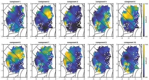
Also, I think it would greatly help the reader to return to PETHs at some point, if possible, to show the response profiles of each identified neuronal subgroup (page 20). To what extent are they similar or different across the cortical areas (for the same neuronal subgroup)?
This is a good idea. We have added a figure to address this question and the related question by R1 (Figure 9-figure supplement 1). In short, given the wide variety of PETHs we observed, there is of course still substantial variation within subpopulation, and some mild but systematic differences in the distribution of what we observe across areas. We now discuss the conclusions from this plot in the Results:
“As a qualitative depiction of the response profiles identified with each subpopulation, we plotted the two highest-likelihood cells for each area/subpopulation combination (Figure 9-figure supplement 1). These examples reveal stereotypy in the subpopulation responses across areas, but also show variation across areas, especially for the two somatomotor subpopulations.”
Specific:
(1) Figure 2B and M&M: the 3D spatial organization of the target locations is not immediately clear. What is the spacing between target locations? What is the 'final azimuthal spacing'?
Added, thanks. The pairwise horizontal distances between targets were between 1.72 and 6 mm apart and the vertical spacing within a column was 1 mm. “Final azimuthal spacing” just referred to the targets being closer together during training and our gradually spacing them apart to their final locations. We have also added some relevant details about the training.
(2) Figure 2C: It would help to have a scale bar (mm).
Added, thanks.
(3) Figure 2C: It would be easier to appreciate the variability of the trajectories across trials to plot an overlay of trajectories to one target only (could be a Supplementary Figure).
The reviewer has a good point: the variability and accuracy of aiming was hard to ascertain from the plot. We experimented with a few options for making this clearer most effectively. We have now added Figure 2-figure supplement 1 that shows in the third subpanel of panel A the finger centroid trajectories for one of the 15 targets highlighted for the mouse shown in Figure 2C, mouse 3. The centroid trajectories for all other mice are shown as well to illustrate similarities and differences across animals as well as the overall variability. As noted elsewhere we have also included an analysis of the variability of the centroid trajectories, showing that reaches to a given target were more similar than reaches to different targets. We think this provides a fuller picture of the behavior and intend to provide still more detail in future work. Thank you for suggesting additional detail here!
(4) Figure 4: It would be nice to also show the amplitude-normalized grand-average PETHs for the different areas.
This is an interesting suggestion. After careful consideration, we think that this analysis is not as effective for depicting overall timing and modulation profiles as the current ones, given the strong amount of target selectivity and response time heterogeneity (now better visible in the revised Figure 4A). When computing the grand mean of all cells within each area, the dominant features distinguishing areas are onset time and response duration. The differences across areas in these two features are better supported by the analyses of Figures 4 and 5 due to the large amount of heterogeneity in responses within each area. We thank the reviewer for encouraging this exploration; more complicated spin-offs will likely inform additional timing analysis in the next paper on these data.
(5) Figure 7C: figure legend - although it is quite self-explanatory, please explicitly indicate which pattern corresponds to the 'Three contour levels (98%, 95%, 90%)'.
We have now added this as a legend on the figure panel itself (here and on similar plots). Thanks for pointing this out.
(6) Figure 8: Is there also an interesting asymmetry between sensory are motor areas, with neurons in sensory areas being more likely associated with motor areas (B and C), whereas neurons in motor regions are less likely to arise from the distribution of sensory areas (dark blue color in frontal regions in D, E, and F)?
This is an interesting observation, but we understand it to be an artifact of colormap scaling. As mentioned above, likelihoods are not well-behaved like probability distributions are: for example, they are not bounded at 1, and their sums over a dataset can have any positive value. The only things that can be interpreted are their relative values. This makes their scaling functionally arbitrary – you’ll notice we used “min likelihood” and “max likelihood” instead of numbers, which would be nearly meaningless – and therefore presents a problem for scaling the colormaps. We don’t know of a principled way around this problem. To deal with it, we simply put the ends of our colormap at the extreme pixel values. It so happens that both the M1 and M2 maps had a handful of neurons in a less-sampled spot at the bottom of M2 that were very low-likelihood, which results in what you noticed. We debated removing those neurons for this purpose, but we had no basis on which to do that kind of manipulation, so we left it as the most honest representation of the data we could produce.
To clarify this, we now mention in the caption “The ends of the colormap were set to the maximum and minimum likelihood values for each map.”
(7) Figure 9B: there are two-time 'S1-hl: 1' indicated at the two bottom rows of the distance matrix. I suppose one of them should be 'S1-tr: 1' instead?
Fixed, thanks for catching it.
(8) Page 20: 'This hinted at a second hypothesis: that some of the 'modes' (groups of neurons) discovered separately in each area might correspond.' ???
We had meant “mode” as in “multimodal”, but it was very unclear. We have rewritten the sentence:
“This hinted at a second hypothesis: that a peak in the multimodal distribution from one area might correspond to a peak in the multimodal distribution of a different area.”
(9) Figure 9S2: Please indicate for which area each map is computed.
The caption was not clear enough about what we were doing here: we fit the GMM on all neurons together, ignoring which area they came from. We have now clarified it in the caption:
“One GMM was fit to the feature vectors of cells from all 5 main areas. Each map plots the likelihood for all cells to each of the three components of this area-agnostic GMM.”
(10) M&M, Subjects and surgical procedures: 'ambient temperature of 71.5 {degree sign}F', please use international units.
Done.
Daarbij worden vooral andere partijen
Ik mis hier en hierboven dat we zelf ook nog steeds standaarden beheren.
bij ontwikkelen
bij ontwikkelen --> bij het ontwikkelen
het
verwijder dit woordje.
gegensopslag
gegensopslag --> gegevensopslag
bij probleemdiagnose
bij probleemdiagnose --> bij o.a. probleemdiagnose
Het is immers ook de bedoeling dat hiermee gecheckt wordt wie wanneer welke informatie heeft opgevraagd zodat oneigenlijk gebruik opgespoord kan worden.
het bericht van BRP gaat naar
het bericht van BRP gaat naar --> het bericht van de BRP gaat bijv. naar
spreken echter
spreken echter --> spreken bij het uitwisselen van gegevens echter
verhuizing verwerkt in het BRP-systeem moet
verhuizing verwerkt in het BRP-systeem moet --> verhuizing, verwerkt in het BRP-systeem, moet
Tip: Je ziet Centric en PinkRoccade Local Government terug in veel domeinen. Dit zijn de twee grootste leveranciers voor de Nederlandse gemeentemarkt. Kennis van hun producten en werkwijze is waardevol.
Ik weet niet of dat hier moet maar misschien wel goed te benoemen dat beide leveranciers (in het verleden) een belangrijke bijdrage hebben geleverd aan onze producten.
eLife Assessment
This study makes a valuable contribution to the understanding of meta-learning and its neural mechanisms by distinguishing two timescales of learning rate adaptation: rapid, within-block reductions and slower, location-specific, meta-learned adjustments. Behavioural data and computational modelling provide convincing evidence that individuals adjust learning rates both rapidly in response to uncertainty and more gradually through meta-learning of environmental statistics. Neuroimaging results indicate that meta-learned learning rates are represented in orbitofrontal cortex, and that prediction errors are encoded across a distributed network including the ventral striatum, where they are modulated by expectations about error magnitude. The manuscript is timely and clearly written and opens the door to future work on how these signals contribute to adaptive behaviour.
Reviewer #1 (Public review):
Summary:
Simoens and colleagues use a continuous estimation task to disentangle learning rate adjustments on shorter and longer timescales. They show that participants rapidly decrease learning rates within a block of trials in a given "location", but that they also adjust learning rates for the very first trial based on information accrued gradually about the statistics of each location, which can be viewed as a form of metalearning. The authors show that the metalearned learning rates are represented in patterns of neural activity in the orbitofrontal cortex, and that prediction errors are represented in a constellation of brain regions including ventral striatum, where they are modulated by expectations about error magnitude to some degree. The work opens the door to future work focusing on how exactly these signals contribute to adaptive behavior.
Strengths:
The authors build on an interesting task design allowing them to distinguish moment-to-moment adjustments in learning rate from slower adjustments in learning rate corresponding to slowly gained knowledge about the statistics of specific "locations". Behavior and computational modeling clearly demonstrate that individuals adjust to environmental statistics in a sort of metalearning. fMRI data reveal representations of interest including those related to adjusted learning rates and their impact on the degree of prediction error encoding in the striatum.
Weaknesses:
It was nice to see that the authors could distinguish differences between the OFC signals that they observed and those in the visual regions based on changes through the session. However, the linkage between these brain activations and a functional role in generating behavior remains somewhat unclear, opening the door for alternative interpretations.
Comments on revised version.
I appreciate the authors responses and they have largely addressed my concerns. I understand the concerns about power with regard to the individual differences/behavioral analyses included in the rebuttal. However, my personal view, which is perhaps a matter of taste, is that the paper would benefit from a description of these results - along with a clear description of why the authors are hesitant to draw a strong interpretation from the negative result.
Reviewer #2 (Public review):
Summary:
Across two experiments, this work presents a novel spatial predictive inference paradigm that facilitates the investigation of meta-learning across multiple environments with distinct statistics, as well as more local learning from sequences of observations within an environment. The authors present behavioral data indicating that people can indeed learn to distinguish between noise levels and calibrate their learning rates accordingly across environments, even on initial trials when revisiting an environment. They complement their behavioral results with computational modeling, further bolstering claims of both local and global adaptation. Additional fMRI results support the role of OFC in this meta-learning process, with central OFC activity reflecting similarity between environments. This similarity emerges over time with task experience. Holistically, this paradigm and these data add to our understanding of how humans dynamically adapt their behavior on different timescales.
Strengths:
The novel paradigm represents a clever and creative expansion of spatial predictive inference tasks. The cover story was well chosen to facilitate an intuitive understanding of both the differences between environments, and the estimation of the mean within environments.
Additionally, the authors present complementary results from two experiments, which strengthens the behavioral findings. This is especially effective as the initial experiment's results were a bit noisy, and the modifications within the second experiment increased both power and the specificity/accuracy of participant predictions. Taken together, the behavioral results provide convincing evidence that participants did distinguish environments based on their underlying statistics and adapted their initial behavior accordingly.
Beyond this, the combination of behavioral results, computational modeling, and neuroimaging enhances the impact of the work. It paints a fuller picture of whether and how humans meta-learn the global statistics of environments, and this is an important direction for the field of adaptive learning.
Weaknesses:
Throughout much of the paper, the authors refer to the distinctions between environments primarily as differences in "initial learning rates" or "environment-specific learning rates." The optimal initial learning rate did indeed differ across environments -- the result of differences in underlying task statistics. These differences in task statistics result in distinct optimal initial learning rates and also vary with aspects of spatial position (e.g. vertical position in the example figure). The authors convincingly show that OFC activity increasingly reflects these variables throughout task experience. Given that these variables vary together, future work will be needed to distinguish whether particular variables drive these dynamics, or whether together they combine to evoke the representational differences.
The current work is also quite suggestive of meaningful individual differences in both local and global adaptive learning, in line with other prior work on predictive inference. This is perhaps underexplored in this data set, but certainly leaves the topic ripe for follow up going forward.
Finally, more information on all clusters that survived multiple comparisons correction would be useful, even in the absence of a priori hypotheses. For instance, there is commentary in the discussion section on the ACC, but this is not mentioned in the results, and it is unclear whether there were other undescribed clusters that survived correction.
Author response:
The following is the authors’ response to the original reviews.
Public Reviews:
Reviewer #1 (Public review):
It was nice to see that the authors could distinguish differences between the OFC signals that they observed and those in the visual regions based on changes through the session. However, the linkage between these brain activations and a functional role in generating behavior was left unexplored. Without further exploration, it is hard to tell exactly what role the signals might be playing, if any, in the behavior of interest.
To link the behavioral with the fMRI data, we now correlated fMRI decoding accuracy with behavioral performance. We studied behavioral performance in two ways: the difference in high versus low noise environment learning rates, and mean accuracy (i.e., absolute prediction error). We correlated both measures with the decodability of the environment in the central OFC. Each correlation was calculated either in the full experiment, or only the second half. However, none of these correlations were significant (all p > .1). Given the difficulty of interpreting this result, and our lack of statistical power for doing individual difference analyses, we decided not to report these analyses in the final paper.
Reviewer 2 (public review):
(1) The authors make the distinction between meta-learned "global" learning rates and within environment learning rate adaptation in response to "local" fluctuations/observations. Though the experimental paradigm is novel, there are certainly links to prior work - for instance, though change point structures don't entail revisiting unique environments, they do require meta-learning from environmental statistics that is distinct from transient local adaptation to prediction errors. This tendency to increase one's learning rate after large prediction errors is appropriate in change point environments, though, as is true in this study, the amount of increase should be dependent on. This represents a similar kind of slower-timescale learning or reuse of more "global" parameters, and can be seen to different extents in prior work. It might benefit readers if the authors were to link the current work to previous research more explicitly to draw clearer connections between the approaches and findings.
We thank the reviewer for their very helpful literature suggestions and now contextualize and discuss our findings in light of relevant literature.
(2) Throughout much of the paper, the authors refer to the distinctions between environments primarily as differences in "initial learning rates" or "environment-specific learning rates." This is particularly prominent when discussing fMRI results. Though the optimal initial learning rate did differ across environments, this was the result of differences in underlying task statistics. It will be important to clarify this throughout the text, because of the confounds between task statistics and initial learning rate (and to some extent, the position on the screen), it is not possible to separate the impact of these specific variables. This is also relevant to understanding the justification for using methods like RSA to test whether brain regions represent task states similarly. If the main hypothesis is that neural activity reflects the (initial) learning rate itself, then a univariate analysis approach would seem more natural.
We agree that task statistics are not the same as differences in learning rates. However, we do not consider this as a confound: The point of the differences in task statistics is exactly to generate differences in learning rates. With our paradigm, we deliberately tried to dissociate variations in learning rate that were induced by learned environmental differences versus local task statistics. We tried to make this dissociation more clear, especially when discussing the fMRI results.
(3) For the neuroimaging results in particular, the specificity of some of the results (e.g. ventral striatum showing an effect of prediction error only in the low noise condition in the second half of task experience, only on the first trial) is a bit surprising. Additional justification of or context for these results would be useful to help readers gauge how expected or surprising these findings are.
We agree some of these findings were unexpected. We now also highlight that while we expected the ventral striatum to be involved in prediction error processing, we had no strong a priori expectations regarding these further modulations by time and environment. We also tried to contextualize these interactions more.
(4) There are some methodological details that are unclear (e.g., how were the positions of the crabs selected relative to the location they emerged from? Looking at Figure 1C, it looks like the crabs spread out unevenly, and that the single position they emerge from is not necessarily at the center of the crab locations.) Additional detail and clarity would help address some unanswered questions (more details below).
We clarified the experimental procedure at several places, and now added a video that helps illustrate the trial timeline better.
Recommendations for the authors:
Reviewer #1 (Recommendations for the authors):
(1) With regards to the primary weakness mentioned above, it would be nice to have some link between the brain signals of interest and upcoming behavior. For example, can you read something out of OFC that enables you to better predict what the participant will do next? Or even better, do so beyond any behavioral variability that is explained by the computational model?
To link the behavioral with the fMRI data, we now correlated fMRI decoding accuracy with behavioral performance. We studied behavioral performance in two ways: the difference in high versus low noise environment learning rates, and mean accuracy (i.e., absolute prediction error). We correlated both measures with the decodability of the environment in the central OFC. Each correlation was calculated either in the full experiment, or only the second half. However, none of these correlations were significant (all p > .1; see plots in Author response image 1). Given the difficulty of interpreting this result, and our lack of statistical power for doing individual difference analyses, we decided not to report this analysis in the paper.
Author response image 1.
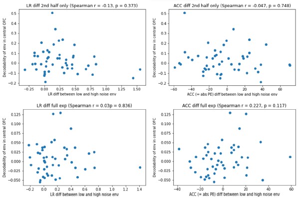
(2) A number of the learning analyses are based on splitting the session into halves. As a first pass, this seems like a reasonable thing to do, but I certainly wonder what the dynamics of the meta-learning actually look like, and it seems like the data collected would be sufficient to gain some insight into those dynamics through some sort of sliding window analysis.
We thank the reviewer for this interesting suggestion, which was also raised by Reviewer 2. We now calculated the learning rate in a sliding window of 20 trials (i.e., trial x to x + 19), and provide revised figures for each experiment separately (Fig. 2E and Fig. 4E, respectively).
(3) The model selection procedures described make sense, but it would still be useful if the authors justified them by showing that they work in synthetic data (ie, generate a confusion matrix). I may be confused about what delta-SE is, but I'm confused about why two models with very different fits have the same value (211) for that metric.
We report model recovery on synthetic data, which yielded model recovery rates of 100%, and added these to our Methods section. To clarify the Reviewer’s second point, ∆SE is the standard error of the difference between a model’s LOOIC and the top ranked model’s LOOIC. There is no one-to-one mapping between the ∆SE and a model’s LOOIC.
(4) Was the central OFC anatomical ROI overlapping with the cluster surviving in the whole brain analysis? I didn't see this mentioned in the text, and it certainly would be important for interpreting the two results together.
The central OFC indeed overlapped with the cluster surviving whole brain analysis, which we report on page 17-18.
(5) The authors found regions that reflected learning rate at the "island presentation" phase of the task - it could be distinguishing this analysis and its meaning from other work that has focused on representations of learning rate at the time of feedback.
We agree that this is an important distinction worth emphasizing. Therefore, we added the following lines to our discussion paragraph:
“Importantly, previous studies examined neural correlates of learning rates during outcome evaluation, where learning rates may be adjusted online as a function of locally experienced prediction errors (e.g., (Behrens et al., 2007; Browning et al., 2015; Nassar et al., 2012). In contrast, our RSA analysis targeted neural activity at island presentation, before any outcome information was available. At this moment, learning rates cannot be updated based on current feedback and instead reflected the retrieval of a previously learned, environment-specific learning-rate settings. This difference reflects our hypothesis that the OFC represents the latent states in a cognitive map of the task (Knudsen & Wallis, 2022; Moneta et al., 2024; Schuck et al., 2018; Wilson et al., 2014), which are expected to activate as soon as the agents can infer which task state it is in. Several studies have identified such “partially observable” task states in the medial OFC (Bradfield et al., 2015; Schuck et al., 2016; Tan et al., 2025; Wimmer & Büchel, 2019), in line with the region identified here (but see e.g., (Ongur & Price, 2000), for important anatomical distinctions between medial and lateral OFC and (Tan et al., 2025) for an example of related functions in lateral OFC). Our finding extends this notion by suggesting a link between OFC and meta learning, wherein meta-learned information becomes encapsulated in task states (Hattori et al., 2023; Moneta et al., 2024).”
(6) "Specifically, it showed a more negative response to larger (location) prediction errors, which is consistent with its documented role in showing a more positive response to more positive reward prediction errors (Calderon et al., 2021) - keeping in mind that being closer to the centre of where the crabs appeared (i.e., smaller location prediction errors) is less negatively or more positively surprising (i.e. smaller negative or larger positive reward prediction errors)."
I found this sentence very hard to parse. Do PE responses in the high noise environment get "compressed" in their representation over time (ie, it takes a larger error to get the same BOLD response)? If so, this relates to claims made in Diederen 2016... but see also Mah 2024 Cell Reports, who fails to see learning rate encoded in DA system in striatum of rodents that appear to adjust their learning rates.
Thank you for pointing to this. We agree that this sentence was hard to parse, and so we now split it in three revised sentences. We also agree with the Reviewer’s interpretation, and would like to thank the Reviewer for their useful literature suggestions which we now added to our discussion.
(7) Figure 7 should use a different color scheme because many of the activations just appear black, and I can't tell whether they are positive or negative. It was also notable in Figure 7A that regions are not visible, including ACC, which is typically thought to encode prediction errors in such paradigms. It would probably be useful for the authors to include a table of all clusters exceeding multiple comparisons correction and to on differences to other work examining absolute prediction errors. ACC does appear on the second trial, which made me wonder whether there were changes in the prediction error coding from first to subsequent trials.
Thank you for pointing this out. We now revised our color scheme which we agree makes it much clearer now. Although the ACC is frequently implicated in prediction error–related signals (e.g., Behrens et al., 2007), models suggest that ACC responses more strongly reflect unsigned prediction errors, surprise, or the need for control and model updating (Alexander & Brown, 2019; Hayden et al., 2011; Silvetti et al., 2018). In our task, ACC activity only emerged on the second trial, when participants had formed an initial estimate and prediction errors could meaningfully signal the need to update internal models or control settings. We now added a to the Discussion highlighting this distinction and relating our findings to this prior work emphasizing prediction errors and control-related signals in ACC.
(8) The authors suggest that fast learning would presumably occur in a neural activation space, whereas slow learning would occur through weight adjustments. This makes sense, but activity-based dynamics have been suggested to do rapid adjustments by encoding a "latent state" though (Razmi 2022 j neurosci) -- and such a latent state has been shown in OFC (Schuck etc)... but here OFC is more implicated in the slow learning. I am curious about whether authors could on this a bit in the discussion.
Thank you for bringing up this interesting question. We can only speculate but a crucial factor is on which level of resolution tasks states operate. On the one hand “detailed” trial-level states are needed that map a specific sensory input onto a specific latent state and its value. Such states would change quickly, possibly through activation dynamics, and are in line with how they have been operationalized in Razmi or Schuck etc. On the other hand, successful task performance also needs “higher level” states that describe entire task phases or full tasks, as in the present experiment. Due to the different speeds of learning, it appears plausible that these would be learned with synaptic changes. We expand on this in the discussion as follows:
“Our finding extends this notion by suggesting a link between OFC and meta learning, wherein meta-learned information becomes encapsulated in task states (Hattori et al., 2023; Moneta et al., 2024). Consistently, OFC has been shown to represent task states (Moneta et al., 2024; Stalnaker et al., 2015; Wilson et al., 2014). While earlier evidence shows that the OFC represents concrete aspects of task states, such as task-relevant stimulus features (Schuck et al., 2016), we hypothesized that the OFC also represents more abstract aspects, such as learned, environment-specific learning rates. Indeed, we showed that the central OFC gradually came to represent these environment-specific learning rates (or the environment-specific statistics that drive them). While previous work speculated that these different levels could have different neural underpinnings (Sharpe et al., 2019), our findings indicate OFC might signal states on multiple levels. This does not imply identical learning dynamics; fast-changing trial-specific states might be learned through activity dynamics, while higher-level contextual states could involve synaptic plasticity.”
(1.9) Also, as a more minor point in the same section, the sentence about blocking synaptic plasticity in OFC sounded interesting, but should have a reference.
Thank you for noticing, we now added the reference (Hattori et al., 2023).
Reviewer #2 (Recommendations for the authors):
(1) Additional links to prior literature: In terms of prior work in which there is something akin to more "global" adaptation, some examples of potentially relevant prior work include:
McGuire, Nassar, Gold, & Kable (2014) Neuron
D'Acremont & Bossaerts (2016) Cerebral Cortex
Lee, Gold, & Kable (2020) Decision
Bakst & McGuire (2021) JEP: General
Bakst & McGuire (2023) Cognition
We would like to thank the reviewer for pointing us to these different literature suggestions which we agree help us contextualize and discuss some of our findings better. We now refer to McGuire et al. (2014) when discussing the fMRI results, and d'Acremont & Bossaerts (2016) when discussing potential alternative strategies in the high noise environment (the Reviewer’s last point). Finally, we integrated the clearly relevant works of Bakst & McGuire (2021; 2023) and Lee et al. (2020) in our discussion of meta-learning different adaptive strategies.
(2) Individual differences: Though not always the focus of work on predictive inference, one common finding has been that there are pronounced individual differences in behavior (see, e.g., coefficients in Figure 2 in Nassar et al. 2019 eLife, or Figure 2 McGuire et al. 2014 Neuron, or Bakst & McGuire 2023 Cognition). There appears to be substantial variability between individuals in your data as well (i.e., Figure 2B, 4B, and the modeling figures). It would be interesting to see some direct exploration of this variability: baseline learning rate appears to differ between participants to a large extent, does their rate of adaptation (across trials within a block) also differ? Does their metalearning occur at different rates (in fact, do some participants not show evidence of appropriate meta-learning at all)?
Relatedly, your computational modeling approach fits the six candidate models hierarchically, and therefore the reported results show the overall best fit for the group. It might be worthwhile to determine whether individuals have different best-fitting models. This could be another way to characterize the variability between individuals.
In concert with this, it could be a useful complement to determine whether either the strength of the OFC neural similarity results or their time course reflects aspects of behavior. Put another way, is it the case that not only does OFC activity and behavior both come to reflect task structure, but that these changes happen to a similar extent and over a similar time course across individuals?
We agree it would be highly interesting to investigate meaningful individual differences in both fast and slow adaptations in learning rate. However, our sample was not set up and is underpowered to conduct such analyses. In response to a similar by Reviewer 1, we did run correlational analyses between differences in learning rate, performance accuracy, and the responsiveness of the OFC. However, none of these analyses yielded a significant effect. We decided to not include these results in the paper, for reasons of statistical power, but we report them in Author response image 1.
(3) fMRI:
(3a) The primary finding in OFC is restricted to the central OFC. The manuscript would benefit from additional explanation regarding this specific subregion.
Thank you for bringing up this important distinction. In the discussion we now clarify as follows:
“This difference reflects our hypothesis that the OFC represents the latent states in a cognitive map of the task (Wilson et al., 2014; Schuck et al. 2018; Knudsen & Wallis, 2022; Moneta et al, 2023), which are expected to activate as soon as the agents can infer which task state it is in. Several studies have identified such “partially observable” task states in the medial OFC (Schuck et al., 2016; Bradfield et al., 2015; Wimmer et al., 2019; Tan et al., 2025), in line with the region identified here (but see e.g., Öngur & Price, 2000, for important anatomical distinctions between medial and lateral OFC and Tan et al., 2025, for an example of related functions in lateral OFC).”
(3b) Though the main clusters visible in Figure 6 are the occipital and OFC clusters, there appear to be others. Did other clusters indeed rise to statistical significance in the whole-brain analysis? If so, is there a reason they aren't included or discussed?
All clusters visible in Figure 6C survived FDR correction. However, we refrained from interpreting these other clusters, because we had no prior hypotheses about them like we did for the OFC.
(3c) Why do you posit that the ventral striatum becomes less sensitive to RPE on the second trial over time? And why is the ventral striatum only sensitive to RPE in the low noise environment generally?
We reasoned the ventral striatum should be more responsive to more positive reward prediction errors. While we further assumed this response could be modulated by both time and environment, we would like to emphasize that we had no specific hypotheses about the direction of this modulation. We now also make this clearer in the manuscript. This being said, we believe both the pattern that its responsiveness to the second trial decreases over time, and the pattern that it was most sensitive to the low noise environment, can be considered fitting with its broader involvement in coding behaviorally relevant reward prediction errors. Namely:
First, we believe that as the participants learn more about the global reward structure of the task, they should obtain a better understanding of the fact that, per round, all crabs always center around a fixed mean. Therefore, the first RPE is most behaviorally relevant, and every later RPE has an exponentially decreasing relevance. As participants obtain more experience with this aspect of the task over time, the VS should show a lower responsiveness to the second RPE over time.
Second, as participants learn more about the local differences between the three different environments, they should learn that especially in the low noise environment, RPEs are most behaviorally informative. That is, in this environment it makes most sense to have a high learning rate and thus let the RPEs substantially inform the placement of the cage on the next trial. Accordingly, participants showed that the ventral striatum was most responsive to RPEs in these environments.
(4) Methods
(4a) This section could generally benefit from some proofreading.
We now proofread the method section.
(4b) The main results text states that 49 participants performed Experiment 1, while the methods section reports 50 participants. Which is correct?
(4c) Following this, on page 8, statistical results are reported with a df = 49 (which would be appropriate only if n=50).
The correct sample size was actually 50, we adjusted the text and degrees of freedom where incorrect accordingly (note: only text is in track changes, but degrees of freedom were also changed accordingly).
(4d) Additionally, I am a bit surprised by the Experiment 1 findings that learning rates on the second trial were significantly different between low and high noise conditions, in that the effect size found using all trials was stronger than both the first half of trials (no significant effect) and the second half (significant but weaker than all trials). Are these all the same type of statistical test? Double-checking the statistics might be worthwhile.
It is not the effect size that is larger across the full experiment, but the t-statistic. This is possible because a t-statistic depends on both effect size and noise estimate, and the latter is smaller with more data.
(4e) The methods and results both state that the five crabs always emerged from one position in the sand. How were the locations of the crabs selected relative to this position? Looking at Figure 1C, it looks like the crabs spread out unevenly, and that the single position they emerge from is not necessarily at the center of the crab locations.
The crabs did indeed spread out evenly. However, we can see how the graphic in Figure 1C can be confusing, as two crabs are shown to be caught, which breaks the symmetry of the dispersion (because some crabs can run away after the even spreading phase, see Methods). We emphasized the even spreading more clearly in the new version of the paper. We think the flow of events will be much clearer with our newly added animation (Video 1).
(4f) The methods section states that the crabs "spread out to cover the same proportion of the screen width as the cage (18.75%)" (page 23). The corresponding visual in Figure 1C appears to show something different.
This looks different because the graphic illustrates the last 500 msec, where crabs can run away (see also response to 4e, and the novel animation that was added).
(4g) Information on the timing of the trials would be useful to include in Figure 1C or similar.
The reader can find this information in the Methods section. We chose not to include it in the caption to avoid information overload.
(4h) The methods section specifies that there was a 3-7s ITI after the first and second trials of each block. How was the ITI selected for each trial? Were there ITIs between the other trials? If so, what were they?
The ITIs were selected from a truncated exponential distribution. This selection was not random, but rather a distribution was carefully constructed for each environment (and event of interest: boat presentation, first trial of each block, second trial of each block) separately to ensure that enough longer ITIs were selected for each environment (and event of interest). Of course, the order in which the ITIs were used across blocks, was random. The same approach was used to determine the duration of the presentation of the boat at the start of each block. There were no ITIs after later trials.
(4i) Please provide a link to the data and analysis materials on OSF in the text.
We now provide a link to the data and analysis materials in our methods section.
(4j) In the methods section, there are some references to information provided "below" (page 26: "The two approaches resulted in different posterior densities (see below) for estimate uncertainties, but in similar posterior densities (see below) for learning rates..."). Where in the paper is this referencing?
We indeed did not detail this further as we considered it not further relevant to our main study, and now removed the references to “below”.
(4k) The methods section specifies using uniform priors between the lower and upper bounds of the relevant parameters. This seems likely to be 0 and 1, but should be listed explicitly.
Thank you for noticing. We now added this to our manuscript.
(4l) For parameter recovery, correlations are provided to indicate effective recovery. These correlations are indeed high and suggest excellent recovery, but correlations wouldn't reveal if there was systematic over- or underestimation occurring. It might be useful to provide some visualizations of the parameters and their estimates to speak to this potential issue.
We now visualize the parameter recovery results in Author response image 2, which show that, indeed, there was a slight underestimation of the decay rates, but not the learning rates. Importantly, our main analyses and results all pertain to the learning rates, and we never made hypotheses or conclusions about the decay rates.
Author response image 2.
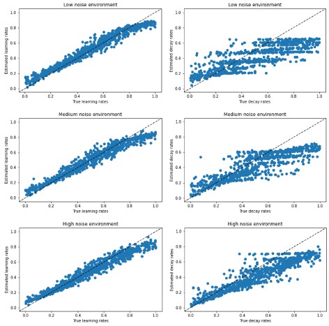
(4m) The methods section ends with a reference to a reward localizer (page 32). This localizer doesn't appear to be mentioned/used elsewhere.
Indeed. We implemented the localizer because we wanted to independently identify reward processing areas. However, this localizer did not succeed in localizing a reward area (no significant results), possibly due to the fact that (1) it was performed by the end of the experiment when participants may have been fatigued, and (2) there was no learning component in this localizer task. For these reasons, we did not use it after all.
(5) Analysis:
(5a) Did you consider fitting a Bai model that only allowed for environment-specific initial learning rates (with a non-environment-specific decay rate)? Given that the data (e.g., Figure 2, Figure 4) seems to support differences in initial learning rate but not necessarily a difference in the rate of change, it might be worthwhile to see whether a model like that fits best.
We now fitted this extra model, which we called the semi-environment-specific Bai model. See Author response tables 1 and 2 for result in experiments 1 and 2, respectively) for the results. This new model has the best (in Experiment 2) and second-to-best (in Experiment 1) LOOIC. In a way, this is not surprising, because the model formulation is entirely based on the data. We think that we can draw the same substantive conclusions with or without this extra model, so for simplicity we did not include this new model in the paper itself.
Author response table 1.
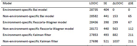
Note. Models are ranked in descending order according to how well they fit the data. LOOIC refers to a model’s approximated expected log pointwise predictive density. Higher values indicate higher out-of-sample predictive fit. SE refers to the standard error of a model’s LOOIC. ∆LOOIC refers to the difference between a model’s LOOIC and the top ranked model’s LOOIC. ∆SE refers to the standard error of the difference between a model’s LOOIC and the top ranked model’s LOOIC.
Author response table 2.
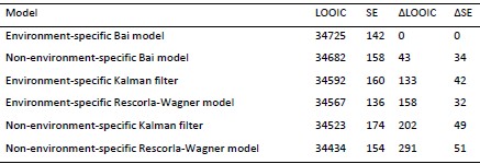
Note. Models are ranked in descending order according to how well they fit the data. LOOIC refers to a model’s approximated expected log pointwise predictive density. Higher values indicate higher out-of-sample predictive fit. SE refers to the standard error of a model’s LOOIC. ∆LOOIC refers to the difference between a model’s LOOIC and the top ranked model’s LOOIC. ∆SE refers to the standard error of the difference between a model’s LOOIC and the top ranked model’s LOOIC.
(5b) If part of the goal is to investigate whether there is a distinct local change in LR between conditions (dependent on prediction errors), then there might be more direct ways of doing so as a complement to the modeling approach. One potential way could be to visualize the LR or change in LR as a function of PE.
We agree that it’s beneficial to use a direct (model-free) approach to represent learning rate as a function of condition; that is also part of our approach. For example, see Figures 2, 4, which shows learning rate as a function of condition, but in a model-free manner. We think learning rate as a function of prediction error is less informative, because the idea is that prediction error can (in Kalman-filter terminology) be indicative of either noise variance or process variance, and participants are able to distinguish between them. This is also why we constructed the conditions in such a way that on the very first trial, prediction errors were on average the same across conditions. The fact that participants did respond appropriately to prediction errors on the very first trial (i.e., larger updates or learning rates in the low noise condition), suggested they are able to assign the prediction error to process variance (in the low noise condition) versus noise variance (in the high noise condition).
(5c) In addition to looking at the evolution of LR across trials within a block separated by task epoch (i.e., Figure 2C-D & Figure 4C-F), the structure of the task would lend itself very nicely to visualizing the evolution of the second trial LR on its own across instances. This could provide additional insight into the meta-learning process.
We thank the reviewer for this interesting suggestion, which was also raised by Reviewer 1. We now calculated the learning rate in a sliding window of 20 trials (i.e., trial x to x + 19), and provide revised figures for each experiment separately (Fig. 2 and 4, respectively).
(6) The environment-specific Bai model appeared to become less good at capturing participant behavior with increased environmental noise. Why do you think this is?
We thank the reviewer for raising this point. In this environment, individual outcomes are considerably less indicative of the latent mean, which may reduce the usefulness of the trial-by-trial, prediction-error–driven learning-rate adjustments that we see in the other environments. Under such extreme conditions of variability, people may rely less on delta-rule updating and more on alternative strategies (D'Acremont & Bossaerts, 2016; Reynders et al., 2026), such as exploratory adjustments or heuristics that are not explicitly captured by the Bai model but also outside the scope of the present paper.
CRIHN
Je l'ajoute, ça ne mange pas de pain, j'en fais partie...
cachets
"contrats" ?
préparation
"tenue" ?
bibliothèque
Idem ?
bibliothèque
Bibliographie ?
Sous
J'ai rédigé un peu ce paragraphe.